The name of the muscles in the hand. All arm muscles: anatomy and proper training
Pumped up embossed arms will decorate any physique. Let's study the anatomy of biceps and triceps, and also see which exercises are most effective for building hand muscles.
Most likely, when you first crossed the threshold of the gym, then immediately started training hands. Both girls and guys want to have slim and beautiful hands, because this is the most visible part of our body. It is impossible to come to the gym and not see at least one person who would perform the lifting for the biceps.
Perhaps you have already trained your arm muscles and did not get the desired result. It's time to learn how to properly and effectively pump up your biceps and triceps. I will tell you about the structure of the muscles, various types of grip, as well as certain exercises that will make a noticeable change in your results.
To more effectively pump up the muscles of the arms, you need to understand how they work. Here are some muscles you should know.
Muscles of the front of the arm
 To pump your impressive biceps, you need to focus on the following 3 muscles: the biceps of the shoulder (biceps), the brachial muscle (brachialis), and the shoulder muscles. It is very important to understand how they differ from each other.
Biceps muscle of shoulder
It has two heads: short and long. The short head originates at the front of the scapula and is attached to the radius (the bone in the composition of the forearm, which ends near the thumb). The long head also originates from the scapula and is attached to the radial bone, but passes a longer way.
Shoulder muscle
It begins in the middle of the humerus and is attached to the ulna (the bone in the composition of the forearm, which ends next to the little finger). It does not participate in the rotational movements of the arm (pronation and supination) due to the fact that it is not attached to the radius. The main function of the humerus is to flex the arm at the elbow.
Shoulder muscle
The muscle of the forearm, which has a great length. It originates from the humerus and is attached to the edge of the radius.

Muscles of the back of the arm
Most people are focused on training biceps, because they look spectacular in the mirror. However, the triceps muscle (triceps) occupies almost 75% of the upper arm, so she also needs to pay a lot of attention. It consists of 3 heads. To pump up the famous "horseshoe", you have to work on all the heads.
Triceps Lateral Head
It originates from the outer surface of the humerus and is attached to the ulnar process of the ulna.
Medial Triceps Head
It originates at the back of the humerus and is attached to the ulna.
Long head
Slightly different from other triceps heads. It starts at the scapula and is attached to the ulna. Due to the fact that it is attached to the shoulder blade, you can isolate it by performing movements with your arms and shoulders. But I will tell about it later.
Bone anatomy
Bones and joints play an important role in arm movement. A clear understanding of their work will help you find the right exercises and pump the target muscles.
Front of the hand
 The biceps workout is affected by 2 main joints. From their position depends on which muscles will be involved in the exercise.
Shoulder joint
It plays an important role in the training of hands, because the long head of the biceps passes through it.
Elbow joint
It also plays an important role, because which muscle will be involved in the work depends on the rotation of the arm. In addition, you will not be able to pump your biceps without bending your elbows.
Back of hand
In training triceps, the same bones and joints play an important role, but for different reasons.
Shoulder joint
The main importance here is the long head of the triceps. To isolate it, you need to raise your arms above your head.
Elbow joint
Arm extension in the elbow is present in almost every triceps exercise. The extension of the arms in the crossover, the extension of the arms in the slope, as well as such basic exercises as a bench press require the extension of the arms in the elbows.
Hand muscle functions
We examined the muscles, joints and bones that are involved in the training of hands. Let's go ahead and see how they work together to make movements. Once you understand the difference between the positions of the shoulders and elbows, you will be able to train your hands much more effectively.
Front of hands
Supine grip (reverse)
Biceps plays an important role in the flexion and external rotation of the arm. Therefore, to more effectively work it out, use the supinating grip.
Parallel grip
When using parallel grip (as in the exercise "Hammers") shoulder muscles are used to the maximum.
Pronirovanny grip (top)
If you use pronounced grip, then reduce the load on the biceps and move it to the brachiocephalic muscles. That is why for their isolation it is recommended to carry out lifting dumbbells with a grip from above.

Back of hands
Arm extension in elbows
When you straighten your arms, you will evenly include all 3 heads of the triceps.
Arm extension in elbows above head
When you press the weight above your head, you will more actively use the long head of the triceps.
Key exercises for training hands
Let's get down to practice. The following exercises will help you build strong and beautiful arms. After watching the video, you may get the impression that these exercises are light, but remember about the intensity of training. You must load your muscles so they grow.
Exercises for the front of the hands
Include these exercises in your biceps training program.
Exercise 1 Lifting a barbell for biceps with an EZ-neck
I like the curved neck more, because it simplifies the exercise. Many people have disproportion in the elbows and shoulders, and this neck allows you to evenly distribute the load on the hands. EZ-neck also reduces the load on the shoulder and elbow joints.

To get the maximum benefit from lifting the barbell for biceps, keep your abdominal muscles and buttocks tense. This will help you lift more weight and protect your lower back. In the lower phase of the exercise, completely unbend your arms, and the upper - fully bend. Use supinating grip to better work the biceps.
Exercise 2 Lifting a rod with an EZ-neck on the Scott bench
In this exercise, use pronounced grip to work out the brachiocephalic muscles. Keep your elbows pressed tightly against the platform of the simulator. Take a short pause in the upper phase of the exercise, and at the bottom fully extend your arms. The range of motion must be complete.

A curved fingerboard provides more insulation, so you cannot lift the same weight that you could lift with a straight neck. Perform this exercise at the end of your workout.
Among other things, this exercise is great for developing the muscles of the forearm. Thus, you simultaneously develop the muscles of the upper and lower parts of the hands.
Exercise 3 Lifting dumbbells with a hammer grip while sitting

This exercise will help to work the shoulder muscles. Use parallel grip (palms facing each other). If you want to complicate the exercise, then slightly rotate the dumbbells (supination) in the upper phase to turn the biceps into work. Keep your abdominal muscles in tension. This will help to comply with the technique and remove the load from the lower back.
Exercises for the back of the hands
Perform these exercises to pump through all 3 heads of the triceps with maximum intensity.
Exercise 1 Press lying narrow grip
In my opinion, this is the best exercise for the triceps. Many of you know that bench press is an exercise for chest muscles. This is true, but in it you unbend your arms, which means you need to work the triceps.

Grasp the neck at shoulder width. If you use too narrow a grip, then in the lower phase of the wrist exercise, too much load will be applied. If the wrists are properly positioned, the knuckles should be facing up. Lower the neck to the chest, tightly pressing your elbows to your body.
Many people believe that all exercises for the hands are insulating (one-joint). But this basic exercise can give the muscles a big load. Here the muscles of the chest are included in the work, which allows you to lift a heavy weight, which means to make the muscles stronger and larger.
Exercise 2 Extending the arms from behind the head with a dumbbell

Raising your arms above your head isolates long tricep heads. This is a simple exercise, but remember that the abdominal muscles must be in tension, and the range of movement was complete. Elbows should remain motionless.
The best result in a scientific approach to training hands
Hands training is more than just a set of isolating exercises at the end of a workout. If you want to build muscle in your arms, then do basic exercises more often.
Do not forget that you work on the muscles of the arms in almost every workout. Biceps and triceps are involved in such exercises as pull-ups and bench press. Exercises for the back, chest and shoulders also indirectly contribute to the development of the muscles of the hands. And if you add to them the above mentioned isolating exercises for the muscles of the arms, then only improve your results.
Build muscle according to the scientific training program
Check out our full six-week workout program. Before you go to the gym and start training, watch the educational videos. Remember that you must combine the work of the muscles with the work of the mind in order to build a beautiful body.
I am sure that the topic that we will address today will not leave anyone indifferent, literally. Why so, you ask? It's very simple, because this topic is the cornerstone of bodybuilding, on which the whole process of building a beautiful and muscular body is based. Without knowing at least the basic principles from this direction, there can be no talk of any significant results in improving the proportions of the body.
Well, I think you have already guessed that the conversation today will be devoted to the topic - the muscles of man.
So, on the agenda consideration of such issues as: the anatomy of human muscles (structure, function and classification), types of muscle fibers and their role in the construction of aesthetically correct body composition. In general, everything you need to know (about muscles) at the initial stage of a newcomer to bodybuilding, we will try to make out today.
Go…
Human anatomy atlas: structure, classification and muscle function
I have long wanted to highlight this question, because I consider him the most important technical and theoretical point of bodybuilding, for you understand that you are trying to build a relief body without knowing (or having a vague idea) about what you have to work with - it's just the height of indecency :).
However, I deliberately postponed consideration of this issue until now, because Immediately it’s very hard to get into the theoretical part - the anatomy and physiology of the muscles, especially when you don’t even know the basics, such as: and the path to bodybuilding success . However, now it does not threaten you, you are already prepared and know these common truths, which means it's time to dig deeper and sort out another important topic.
So every newbie (and not only him) bodybuilding is simply obliged to know as much as possible about the muscles, their functions and structure, because in this way he will achieve much more impressive results than his fellow iron. Agree, want to build muscle and do not know the elementary basis of their physiology, smacks of marasmus. Although, I will tell you in secret, when you begin to ask the gym regulars, what kind of muscle they swing or where is brachyalis, they make such eyes as if they don’t understand what it is all about. Well, okay, closer to the body.
So, a human muscle is an organ of the body (soft tissue) consisting of muscle fibers that can contract under the influence of nerve impulses and provide the basic functions of the human body: movement, breathing, nutrition, resistance to stress, etc.
From this definition, it is immediately possible to make the obvious conclusion that the brain and muscles are interconnected. (what he will order will be fulfilled)hence from the brain-muscle ligament (or rather, from the speed of the neuromuscular reaction) depends on the efficiency of pumping muscles. And one more conclusion arising from the previous statement is that we need to work not only on pumping the muscles, but also on reducing the response time of the neuromuscular response, i.e. be friends with your head.
The muscle consists of striated, cross-striped bundles of muscle fibers running parallel to each other, which are connected by connective tissue into bundles of the first order. Several such beams are connected and form second-order beams, etc. All these muscle bundles are combined with a special sheath, making up the muscular abdomen (see image).

When a muscle contracts (under the influence of nerve impulses)It distinguishes between the "actively contracting" part - the abdomen and the passive part, with the help of which it is attached to the bones - the tendon. Generally speaking, skeletal muscle is a complex structure consisting of striated muscle tissue, of various types: connective (tendon) and nervous (muscle nerves) tissues, endothelium and smooth muscle fibers (vessels).
Well, impressive?
From all this it is worth remembering that the striated muscle tissue, whose property (contractility) determines the function of the muscle as a contraction organ, is predominant in the muscle structure. The human body consists of various muscle groups - a complex of several muscles that performs the same motor function. When performing similar exercises in the same movement, almost all muscles from the same muscle group are usually involved in the work.
Let us turn to the classification, and the easiest way to present it is in the form of the following pivot table (see table).

Let us analyze and analyze some classifiers.
№1. In form
There are short, long and wide muscles. The habitat of the long muscles (mostly) is the limbs, with their help, exercises with a full amplitude of movement are performed. For example, lifting a barbell for biceps is one such exercise in which this type of muscle works.
Long muscles are tadpoles, because consist of three parts: the beginning of the muscle - the head, the middle part - the abdomen of the muscle, the end of the muscle - the tail. They are similar in shape to spindles, tendons have the appearance of a narrow ribbon. Vivid representatives of the family of long muscles are those that end in “-ceps”: biceps (two-headed), triceps (three-headed) muscles of the shoulder, quadriceps (see image).

Also, the long ones include those that are formed as a result of the fusion of muscles of different origin, usually multi-abdominal muscles that have 2 or more abdomen. A bright representative is the abdominal and other abdominal muscles.
The wide muscles are located mainly on the body and have an extended tendon. Vivid representatives of the broad muscle family are, for example: superficial muscles of the back and chest. Short muscles are similar in shape to either long or wide muscles, but are much smaller in size relative to their fellows.
Also, there are other forms of muscles: round, deltoid, soleus, gastrocnemius, etc.
№2. In the direction of the fibers
There are:
- Straight-parallel;
- Skew;
- Transverse;
- Circular.
Straight-parallel muscles can significantly shorten with contraction, which provides greater amplitude and an increased trajectory of movement. Oblique muscles are inferior in their ability to shorten, but due to the fact that they are more numerous, they are able to develop greater force.
And in confirmation of these words - an example from a training life.
Often, due to ignorance of the anatomical features of the muscles, people try to give that muscle load that the latter, in principle, is not designed by nature. For example, due to the fact that the biceps refers to the type of straight / parallel / long muscle (by classification), he cannot a priori take that weight, which the oblique muscle will pull, but the latter, by virtue of its numerous fibers, will easily develop a greater force (traction) force. Well, to understand this, you need to know the basics of physiology, muscle anatomy and understand the principles of their work. (let's talk about the last further).
So, the following - transverse and circular muscles. The cross-beams are similar to oblique and perform similar types of work in many respects, while the circular ones are different from them in that they are located around the orifices of the body. (e.g. mouth muscle) and by their contraction narrow them. The second name of these muscles - compressors or sphincters, the most popular of them, is the sphincter of the “fifth point”.
Number 3. Patterns of muscle location
It would be naive to believe that there are no patterns in the distribution of muscles throughout the body, of course they are, and they sound like this:
- According to the structure of the body and taking into account the principle of bilateral symmetry, the muscles are paired or consist of two symmetrical halves.
And glory to God, that symmetrical, and then imagine a face of two different halves, that is still a picture. Although after a series of holidays, some people can see a clear bias, for example, the front halves :); - The human body and, in particular, its body, consists mainly of segments (separate independent elements) muscle Those. this is not some kind of solid general layer (although the wide abdominal muscles of this type), and there is a clear (sometimes conditional) division into sections, for example, the rectus abdominis muscle can be divided into upper and lower sections;
- Muscles are located at the shortest distance between their points of attachment. The movement produced by the muscle is performed in a straight line, therefore, knowing the points of attachment of the muscle, and the fact that the movable part is attracted to the stationary one, it is possible to determine in advance the direction of movement and muscle function;
- The muscles, spreading over, cross at right angle the axis in the joint around which they produce their movement.
So, we have dealt with the classifiers of the muscles and the geography of their location, now let's consider some technical issues in pumping high-quality muscle mass, to which few people pay their attention or do not take them into account at all.
Human muscles: an introduction to mechanics. How that works and works.
Cell structure and muscle contraction
We all know that any living organism consists of cells — the smallest structural unit. So, the structural unit for the muscles is myofibrils - the thinnest threads running along from one end of the muscle fiber to the other.
Since muscles (largely) perform a contractile function, then active elements — protofibrils in the form of actin proteins — are used to ensure this activity. (long and thin fibers) and myosin (short and twice as thick as actin fibers). In different types of muscles, for example, in smooth and skeletal, protofibrils are located differently. Thus, in the smooth, the latter are arranged in a disordered manner and, mainly, along the periphery of the inner surface of the myofibrils. In skeletal muscles, actin and myosin are strictly ordered and occupy their entire internal cavity.
One can observe such a picture - places where actin fibers partially enter between the fibers of myosin look like dark stripes, and other particles appear light, therefore such myofibrils are called cross-streaked.
In general, in the most general form, the mechanism of muscle contraction is as follows: when muscle is contracted, myosin fiber, using energy ATP (adenosine triphosphate acid) moving along the actin fibers, thereby providing the main function of the muscles (see image).

Myosin in this process plays the role of the enzyme adenosine triphosphatase and promotes the breakdown ATPand release of energy.
Summarizing all the above, you should remember that smooth muscles (due to its structure) shrink relatively slowly (from a few seconds to 1-4 minutes)whereas the striped muscles are able to contract very quickly (for a split second). Keep this in mind when working with this or that type of muscle.
The principle of the muscles: the elements of biomechanics
In this subsection, it is appropriate to recall the saying: “What kind of driver does not like to drive fast?”; In relation to bodybuilding, you can say this: “What newcomer does not want to pump a large, shaped body?” But this will help (including) knowledge of the principles of biomechanics of the muscles, i.e. It is important to understand what processes and, most importantly, how they proceed when working with iron.
Agree, there is something like this, when you, while picking up a dumbbell, do not just do the memorized exercise on autopilot, but understand what happens in your muscles at that moment. Understanding these issues will advance you towards the quality and quantity of muscles on your body.
So, the main property of muscle tissue (as we have repeatedly said) - contractility. This process is a shortening of the muscle and the convergence of the two points to which it is attached, with the movable point being attracted to the fixed one.
By acting in this way, the muscle not only produces traction. (moves the load)but also does mechanical work. And now attention! The strength of the muscle depends on the number of muscle fibers that make up its composition, and is determined by the area of the physiological cross-section, that is, the area of the incision in the place through which all the fibers of the muscle pass. Thus, the length of the muscle affects the amount of its contraction.
In a sense, you can compare bones moving in the joints under the influence of muscles with mechanical levers for moving objects of varying degrees of severity.
Thus, it turns out that the farther from the place of support muscles will be attached, the more effective it is, because due to the increase in the lever arm, their strength can be better used - theoretical mechanics, 3 Institute course, comrades dear.
If we proceed from the “lever concept”, then we can distinguish strong muscles — attached far from the fulcrum point and deft attached — attached near it. First (e.g. soleus), do a better job of static nature. They are characterized by a large surface of their origin and their place of attachment is close to the point of gravity. Also, strong muscles are richer in blood vessels and muscle pigment (myoglobin), their color is darker, so they are called red.
These muscles do not tire for a long time and during work they show great strength with a slight tension. However, good power indicators affect the speed and scope of their movement while reducing, which are small. It can be said that strong muscles are certain supporting points of the whole human musculature.
The situation with deft muscles is quite different. They (e.g. biceps muscle of thigh), characterized by long, usually parallel fibers, with a small area of the beginning and attachment, as well as fewer blood vessels, so they are called white muscles.
These muscles are distinguished by the speed of contraction and, working with great tension, they tire quickly enough. Although the white muscles are inferior in red, they are able to perform various movements quickly and in an explosive manner. More often you can find the name of these fibers: slow (red) and fast (white) or fibers of the first ( I) and the second ( IItypes (see image).

In the human body, the muscles contain both red fibers - of a static type, and white fibers - of a dynamic one. Basically, 3/4 people of the planet have a percentage of red and white fibers such: 60/40 i.e. The former prevail, but professional athletes are outside these parameters. Do sprinters in general to 90% leg muscles are rapidly contracting, and I thought what they were scratching right from the start :).
Of course, with age (as well as the magnitude of the load) the ratio of white and red fibers varies.
And also, if during your life you don’t lift anything heavier than a tablespoon or TV remote, then your muscle cells are updated every 7 - 15 days If you work out in the gym, work with iron, then the update process is accelerated, because any exercise (and especially with burdens) provoke the occurrence of muscle fiber breaks, thereby stimulating the growth of new cells.
Note:
Besides the fact that every muscle has a “beginning” (which usually coincides with the fulcrum) and the attachment, its movable and fixed parts may be interchanged.
In general, a large number of exercises in bodybuilding, where movements are performed in opposite directions (flexion / extension - press, adduction / abduction - work with dumbbells, etc.) requires the participation of at least two mutually located muscles. Such human muscles acting in mutually opposite directions are called antagonists. It is thanks to them that smooth movements are ensured. So, if there are antagonists, then there are also opposing muscles, the resultant of which runs in the same direction and they are called agonists, or synergists. It should be understood that the same muscles can act as both synergists and antagonists.
Now consider the issues of involvement of muscles in the work and the processes of their blood supply.
Muscle work: neuromuscular activity
Muscle is a soft and elastic body that can be stretched by external forces. Therefore, as soon as the stretching process begins, excitation occurs in its receptors, reaching along the nerve fibers CNSwhich then returns back to the muscle, causing its tension (opposed to stretching).
Any work characterizes her. Efficiency. The efficiency of the work of the muscles is the developed “muscle-strength” thrust and the amplitude (range) of the movement of the latter. By the force of thrust, we mean the amount of tension that develops in a muscle when it is excited. The thrust force is in direct proportion to the number and direction of the fibers. The muscle is stronger, the more muscle fibers it contains. However, counting the latter is almost impossible. Therefore, there is such a thing as the physiological diameter of the muscle, and that is what determines the strength of the latter.
Note:
Physiological diameter - the cross-sectional area of the muscle in the plane perpendicular to the length of all its fibers. Each square centimeter of physiological width of the muscle can withstand an average 10 kg of cargo.
Also, the parameter characterizing the work of the muscle is the range of movement, which depends on the nature of the bone skeleton, on the length of the muscular abdomen and “lever arm”, as well as on the muscle itself.
It is worth mentioning about another important point relating to the force developed by the muscle. Of great importance for the force of thrust is the degree of excitation of the muscle. The stronger the stimulating effect of the nervous system, the greater the amount of muscle fibers that captures the excitement, the greater the force of thrust. The influence of the nervous system, like the circulatory system, depends on the general condition of the body, the type of higher nervous activity, etc.
Trophy and innervation of muscles
In addition to the fact that the work of muscles, as well as other organs, is regulated by the nervous system, the bloodstream also contributes to the management.
As we understood (we understood, right? :)), the muscles simply perform a tremendous amount of work, and the metabolic processes in them are very active, so it is not surprising that they have an extensive network of blood vessels, through which blood brings nutrients and oxygen to them, and removes metabolic products.
It is worth noting that, of course, not all muscles are equally equipped with blood vessels. For example, those that work almost “day and night” (diaphragm, heart, etc.), have an extensive circulatory system. Those that are not included in the work for a short period of time do not have such a network. (for example, biceps, rectus abdominis, etc.). Nervous ending of any muscle receptors or effectors, which are located wherever possible: in the muscle tissue, tendons, etc.
From the lips of many successful bodybuilders you can hear the following words: “in exercises you need to feel a muscle”, this feeling is achieved by perceiving the receptors of a certain degree of muscle contraction / stretching. Thus, the nerve fibers, as the electrical wires transmitted signals from the brain to the muscles and vice versa (see image).

Effectors are also nerve endings that transmit arousal. (coming from the nerve center as a response to a change in state perceived by the receptors) muscle Do you understand what you said? :)
In general, I think you understand that the neuro-sympathetic mechanism plays a significant role in pumping high-quality muscle mass.
A few words about the development and not the development of muscles
It is so conceived by nature that the human body is created for various kinds of activity through the work of muscles.
Modern realities prove that we are increasingly forgetting to use the latest in our daily life and are heavier than the remote from the TV trying not to take anything. All this leads ultimately to muscle atrophy and loss of their performance. Only constant power loads, workouts in the gym, and fitness classes will allow you to include the muscles designated for this in your direct activity. As a result, all this will lead to an increase in volume, an increase in muscle strength, and a general physical development of the whole organism.
Uff-f, like, all lit up what he wanted. Are you tired? The fact is, but how much useful and important have you learned about these very muscles.
All you need to know about muscle
I would like to finish criminal summary a summary table, so to speak, summing up the totals of all that was said here (a lot has been said here, believe me), so that you all finally settled on the shelves.
So, here are the main points that need to be learned:
- Study the information on all groups of muscles of the human body in more detail, in order to understand how to work with them more effectively;
- Feel your whole muscle working as you do the exercises;
- Remember about the types of muscle fibers: white and red, and engage in the work of both types of fibers to achieve the desired volume of muscles;
- Remember that muscle strength depends on the number of muscle fibers that make up its structure, and increase them;
- Work with muscle antagonists. (acting in mutually opposite directions),so and synergists (acting in one direction);
- Stimulate your nervous system in the approaches when working with weights, in order to involve a non-illusory large number of muscle fibers;
- Remember that an extensive circulatory system is important for the full trophic (nutrition) of the muscles, so if you do not deny yourself the pleasure - “pitch”, think, is it worth it?
- Do not run your muscles, they should work whenever possible.
Now for sure.
Well, my dear readers, we did such a large amount of work (you and I), namely, we dealt with human anatomy issues, examined their classification, learned something about cell structure and muscle contraction, and much more. I would like to add that soon we will look at a person’s muscular atlas in a section, with specific examples of muscle groups, also in detail and in detail. However, this is another story.
On this all, stay with the project “”, subscribe for updates and may the force be with you!
Ps. As always, I will be glad to receive your comments, questions, greetings, and so on. Share the article with others, and most importantly, come again, because here you are always welcome!
Hello friends! Muscles of the arms: the anatomy of this muscle group is very interesting, and it will be useful for many to learn it in order to understand where and how to direct their efforts in the training process.
Despite the fact that I am not a huge bodybuilder even now (I am only 91-92 kg of “dirty” weight now), I have never really had problems with the formation of arm muscles.
They grew by themselves, honestly.
Well, not that I lay up the fifth point on the couch, and my hands increased by themselves, no.
Simply, without doing anything special in the hall (the usual standard, illiterate workouts of 99% of beginners, such as “pull more, push further”), yes, and there wasn’t anything supernatural, even in this situation, my hands didn’t seem to be thin branches.
They somehow respond well to the load themselves. For any workout, at any intensity.
Later, when I began to closely study all the subtleties of the training process, physiology, human anatomy, then it became clear to me why this was happening and happening.
I will tell you all the very cool training exercises in the next article about the powerful training of hands, and today we will focus on ANATOMY, so as not to interfere with kissel and mashed potatoes, so to speak.
Arm Muscles: Anatomy
The muscles of the arms are divided into:
- The muscles of the shoulder.
- The muscles of the forearm.
The muscles of the shoulder, in turn, are divided into:
- Flexors (front group).
- The extensors (rear group).
The group BENDERS includes:
- Shoulder muscle.
- Biceps muscle of shoulder.
- Klyuvovidno-humeral muscle.
The group of unbrokers includes:
- The triceps muscle of the shoulder (triceps).
- Elbow muscle.
We will not disassemble the muscles of the forearm today, because there will be a separate, detailed article about them.
As the muscles of the shoulder, as we see, nothing complicated.
Let's look at a little more.
The muscles of the hands that interest us most
I never understood why focusing on small muscle groups, if underdeveloped large.
Why build a veranda when a house has not yet been built?
Indeed, a much greater release of anabolic hormones is given by training of large muscle groups.
That is why the greatest interest for us among all the muscles of the hands will play only (by decreasing their size):
- Triceps (triceps muscle).
- Brachialis (shoulder muscle).
- a large "horseshoe-shaped" arm muscle, consisting of three heads.
From the side, triceps, indeed, looks like a horseshoe.
- Long triceps head (internal) - attached to the back of the blade. It requires maximum retraction of the arm (ideally, an additional shoulder joint should be included in the work, since the long head is involved in its extension).
- Lateral head of the triceps + Medial head of the triceps- attached to the humerus. They participate only in unraveling the forearm.
All three heads on the other hand are attached in the area of the elbow joint, therefore, no matter what exercise for triceps you use, you will always include ALL THREE HEADS in the work!
But the degree of involvement in the work of all heads will be different, depending on the mechanics of movement.
On the side of the elbow, there is also a tendon that unites and attaches to the bone ALL three triceps bundles. This is purely a feature of GENETICS, and the workouts are not corrected in any way.
- If you have it short, then the triceps is more “smeared” on the back of the hands, it turns out to be more massive.
- If you have a long tendon, the triceps is more PEAK, short.
There is such a pattern (but not the rule!), If you:
- or mesomorph = triceps more massive, long.
- = triceps less massive, but more peak, aesthetic.
Triceps Functions: extension of the forearm in the elbow joint (long, lateral and medial heads), extension of the shoulder and bringing it to the body (only the long head)
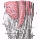
Biceps
Biceps (biceps muscle of shoulder) is a large muscle, clearly visible on the front surface of the shoulder, consisting of two heads (“bi” = two).
- Long head (long tendon, but a small part of the biceps): located from the outer part of the biceps.
- Short head (short tendon, but most of the biceps): located on the inside of the arm.
Both heads of the biceps are attached to one tendon, which, in turn, secures the biceps to the elbow joint. There is a similar situation with triceps, remember?
But, since the ligament is attached to us not exactly exactly (slightly inward, to the lateral part of the forearm), the biceps can not only bend the arm in the joint, but also SUPPORT (unfold) it towards the thumb.
With the development of the internal biceps head, no one has any particular problems, since it grows from almost any flexion.
But the external head lags behind the vast majority.
It so happened that anatomically, the external head is attached to the shoulder joint in the upper part, therefore, to engage it in work, you MUST RETURN LOCTORS BACK (which almost no one does). This will stretch it, purely from a mechanical point of view, and make it work.
A lot of interesting pieces will be in the next article, about the training of hands, but for now let's focus on these moments of the biceps anatomy.
Biceps Functions: flexion of the shoulder in the shoulder joint, flexion of the forearm at the elbow joint, supination of the forearm outwards.
Brahialis
Brahialis (shoulder muscle) - a muscle that is UNDER BICEPSOM (as a lining), but performs directly BENDING (does not participate in the process of hand turning, because it is attached exactly exactly, and not laterally, like a biceps).
This is a very important point, it is the brachial muscle that allows you in the gym to “pukanistye” weight on in any flexion, it is she who performs 65-70% of the work, and not biceps at all, as is commonly believed.
It is fastened strictly exactly to the bone, and not to the side, as the biceps, so the motion vector is concentrated on flexion in the elbow joint.
I will look at the details of brachialis training in detail in the next article. It is very important to develop it, because this muscle, as it were, PUSHES the biceps out (the more brachialis, the stronger the biceps stick out).
Muscles are needed not only to be proud of their relief, but also to make movements. They represent the basis of the musculoskeletal system and serve as the muscles of a person to perform any voluntary and involuntary actions. To know the anatomy of muscle tissue and their types should any professional athlete who wants to quickly and efficiently pumped. After all, it depends on the preparation of the training complex and the distribution of the load.
A person has these main types of muscles:
- Not streaked. They are involuntary muscles and they are also called smooth. Not striated muscles contract rather slowly and in this position they are able to stay for a long period of time without spending a lot of effort. This type of muscle activity is called tonic. Involuntary muscles are subordinated only to the vegetative part of the nervous system; therefore, human consciousness does not affect them in any way. There is such muscle tissue in the skin organs, blood vessels, etc .;
- Squared (skeletal). Such a group of human muscles is arbitrary and is a formation in the form of a cylinder. Their fibers are intertwined and are connected by tendons. The size of the skeletal muscles usually varies from 5-6 mm to 15 cm and more, and their average diameter is 40-60 microns. The striated muscular tissue is contained in skeletal muscles, as well as in the esophagus, pharynx, etc. Such muscles contract mainly intensively and arbitrarily, but at the same time they get tired quickly. This process is called tetanic. Involuntary actions are characteristic only of the muscles of the pharynx of the larynx, etc.
You can look at the structure of human muscles on this scheme:
Muscle movements are usually of the following types:
- Dynamics. It represents the movement of a body or individual limbs to perform certain actions;
- Statics. This kind of movement is designed to fix the body in one position, for example, sitting at a computer.
An athlete must know his body, so the name of a person’s muscles and all their anatomical features, including location, must be known to him, and this can be understood with the help of this image:

Focusing on this scheme, a picture of the muscles of a person with the names you can understand how they are called and what they are, for example, on the legs and arms. Such knowledge will not prevent not only medical staff, but also ordinary people and professional athletes.
Muscle tissue support
The musculoskeletal system includes not only the muscles of the human body, but also the bones, since it is on the skeleton that all tissues are held. You can see its structure in this image:

The moving component of the skeleton are the joints. Their ends are tightly connected in the articular bags. Due to the work of the joints in combination with muscle contraction all movements occur.
Deltoid muscle tissue
Deltas are one of the most visible muscle groups in humans. After all, volumetric shoulders are practically always looked at during visual contact with a person. For athletes involved in bodybuilding, the delta is considered an extremely important component of an ideal figure. You can pump them with the help of swings with dumbbells, bench press and barbell thrust. You can study the structure of this muscle group and its functions in this image:

Chest muscles
To achieve the growth of the pectoral muscles is not easy, because for this you need to try a lot and build the training process correctly. Such a group of muscles emphasizes the expressiveness of the relief of the athlete and his masculinity. In size, the pectoral muscles are inferior to many others, for example, the muscle tissue of the back and legs. However, they are actually half the front part of the torso, so they immediately catch the eye. For pumping are used mainly push-ups and exercises on the bench. This muscle group is divided into large and small pectoral muscle. You can look at the functions and their structure in this picture:

Spinal muscular group
The back is considered a definite stabilizer of the entire human body and the most extensive muscle group. It includes a trapezoid, as well as a diamond, the broadest and many other muscles. They can be pumped through the use of certain exercises that can load certain muscle groups. Well helps tightening with a wide grip, as well as the press and pull in the slope. You can see the structure of the muscle tissue of the human back in this image:

Abdominal muscles
Blown press looks good in both women and men. On the convex cubes almost immediately pay attention. However, to achieve them is extremely difficult, because this area is a problem for many people. If the abdominal muscles are tightened, then it is good not only from an aesthetic point of view, but also for health, since a person will be able to bear more exertion and various diseases will not arise. The basis for pumping this muscle group is a press on the floor and crossbar, as well as various types of twisting. Look at the anatomical features of the abdominal muscles in humans can be in this image: 
Muscles on hand
The arm muscles are predominantly represented by the biceps and triceps, but also brachialis and brachio-muscular tissue. They serve to bend and rotate the arm, so exercises with dumbbells and a barbell are ideal for pumping. Pull-ups and push-ups will fit no worse if there is no inventory. You can look at their structure on this image:

Muscle tissue of the legs
The legs are a large muscular group, the basis of which are the muscles of the lower leg, thighs and buttocks. Due to them, a person can perform certain movements with their feet and walk. Each of these muscle tissue has its own characteristics of pumping and you need to study their anatomical features before training. You can see the structure of a person’s buttocks in this image:

The structure of the thigh also has its own anatomical features, which can be seen below:

The muscles of the human leg are clearly visible in this picture:

Focusing on the anatomy of the muscles of the legs in the picture, anyone can choose for themselves a set of exercises that will load the problem muscle group. The most popular exercises are squats and leg outs. Work in the gym also will not be superfluous.
Muscles in humans are arranged in such a way as to move correctly and harmoniously and perform certain movements. Knowing their structure and functions, you can focus on a specific muscle group to pump it up. In this case, it will be possible to achieve impressive relief and not leave a single problem area.
No matter what a person does - walk, run, drive a car, dig the ground, write, - he performs all his actions with the help of skeletal muscles. These muscles are the active part of the musculoskeletal system. They hold the body in an upright position, allow you to take a variety of poses. The abdominal muscles support and protect the internal organs, i.e. perform supporting and protective functions. Muscles are part of the walls of the chest and abdominal cavities, the walls of the pharynx, provide movement of the eyeballs, auditory ossicles, breathing and swallowing movements. This is only a partial list of skeletal muscle functions.
Therefore, it is not surprising that the skeletal muscle mass in an adult is 30-35% of body weight. In humans, more than 600 skeletal muscles, they are formed by striated muscle tissue.
1 - Scheme of the structure of muscle fibers:
a - myofibril
2 - Scheme of the structure of the myofibrils:
a - shell
b - myosin
d - bridge between them
d - nerve fiber
Each muscle consists of parallel bundles of striated muscle fibers. Each bunch is dressed in a sheath. And all the muscle outside is covered with a thin connective tissue sheath that protects the delicate muscle tissue. Each muscle fiber also has a thin sheath on the outside, and inside it there are numerous thin contractile filaments — myofibrils and a large number of nuclei. Myofibrils, in turn, consist of the thinnest threads of two types - thick (protein molecules of myosin) and thin (actin protein). Since they are formed by different types of protein, alternating dark and light bands are visible under the microscope. Hence the name of skeletal muscle tissue - striated. In humans, the skeletal muscles consist of two types of fibers - red and white. They differ in the composition and number of myofibrils, and most importantly - the features of contraction. The so-called white muscle fibers contract quickly, but quickly and get tired; red fibers shrink more slowly, but may remain in a reduced state for a long time. Depending on the function of the muscles, certain types of fibers predominate in them. Muscles do a great job, so they are rich in blood vessels, through which blood supplies them with oxygen, nutrients, makes metabolic products. The muscles are attached to the bones with the help of inextensible tendons, which grow together with the periosteum. Usually, the muscles at one end are attached above, and the other below the joint. With this attachment, muscle contraction drives the bones in the joints.
Depending on the location of the muscle can be divided into the following large groups: the muscles of the head and neck, the muscles of the body and the muscles of the limbs.

1. Superficial flexor.
2. Pectoralis major muscle.
3. Deltoid muscle.
4. Biceps muscle of the shoulder.
5. Fibrous plate.
6. Beam finger flexor.
7. Anterior gear muscle.
8. Quadriceps.
9. Tailor's thigh muscle.
10. Anterior tibial muscle.
11. The cruciform muscle.
12. The calf muscle.
13. Biceps muscle.
14. The gluteus maximus muscle.
15. The external oblique muscle of the abdomen.
16. The triceps muscle of the shoulder.
17. Biceps of thigh.
18. Deltoid muscle.
19. Trapezius muscle.
20. Subostine muscle.
21. Diamond-shaped muscle.
22. The biceps muscle of the shoulder.
The muscles of the body include the muscles of the back, chest and abdomen. There are superficial muscles of the back (trapezius, latissimus, etc.) and deep muscles of the back. The superficial muscles of the back provide movement of the limbs and partly of the head and neck; deep muscles are located between the vertebrae and the ribs and, with their contraction, cause extension and rotation of the spine and maintain an upright posture.
The chest muscles are divided into the upper limbs attached to the bones (large and small pectoral muscles, anterior gear, etc.), carrying out the movement of the upper extremity, and the actual muscles of the breast (large and small pectoral muscles, front toothed, etc.). thereby ensuring the act of breathing. This group of muscles also includes the diaphragm, which is located on the border of the chest and abdominal cavity. The diaphragm is a respiratory muscle. When contracted, it lowers, its dome flattens (the chest volume increases — inhalation occurs), when relaxed, it rises and takes the shape of a dome (the chest volume decreases — exhalation occurs). In the diaphragm, there are three holes - for the esophagus, aorta and inferior vena cava.
The muscles of the upper limb are divided into the muscles of the shoulder girdle and the free upper limb. The muscles of the shoulder girdle (deltoid, etc.) provide the movement of the arm in the area of the shoulder joint and the movement of the scapula. The muscles of the free upper limb contain the muscles of the shoulder (the anterior group of flexor muscles in the shoulder and elbow joint — the biceps of the shoulder, etc.); the muscles of the forearm are also divided into two groups (front - flexors of the hand and fingers, back - extensors); muscles of the hand provide a variety of finger movements.
The muscles of the lower limbs are divided into the muscles of the pelvis and the muscles of the free lower limbs (muscles of the thigh, lower leg, foot). The pelvic muscles include the ileo-lumbar, large, medium and small gluteus, etc. They provide flexion and extension in the hip joint, as well as maintaining the vertical position of the body. Three groups of muscles are distinguished on the thigh: the anterior (quadriceps of the thigh and others unbend the lower leg and bend the thigh), back (the biceps of the thigh and others unbend the lower leg and bend the hip) and the inner group of muscles that bring the thigh to the midline of the body and bend the hip joint . There are also three groups of muscles on the lower legs: the anterior (unbend the fingers and foot), the posterior (gastrocnemius, soleus, etc.), bend the foot and toes, and the external (bend and retract the foot).
Among the neck muscles, the superficial, middle (muscles of the hyoid bone) and deep groups are distinguished. Of the superficial, the largest sternocleidomastoid muscle tilts back and turns its head to the side. The muscles located above the hyoid bone, form the lower wall of the oral cavity and lower the lower jaw. The muscles located below the hyoid bone, lower the hyoid bone and ensure the mobility of the cortical cartilage. The deep muscles in the neck tilt or turn their heads and lift the first and second ribs, acting as the respiratory muscles.
The muscles of the head make up three groups of muscles: the chewing, facial and voluntary muscles of the internal organs of the head (soft palate, tongue, eyes, middle ear). Chewing muscles drive the lower jaw. Mimic muscles are attached at one end to the skin, the other - to the bone (frontal, buccal, zygomatic, etc.) or only to the skin (circular muscle of the mouth). By shortening, they change the facial expression, participate in the closure and expansion of the openings of the face (eye sockets, mouth, nostrils), provide mobility of the cheeks, lips, nostrils.

Muscles, contracting or straining, produce work. It can be expressed in the movement of the body or its parts. Such work is accomplished with lifting weights, walking, and running. This is a dynamic job. When holding the body parts in a certain position, holding the load, standing, maintaining the posture, static work is performed. The same muscles can perform both dynamic and static work. By contracting the muscles, they move the bones in motion, acting on them as if on levers. The bones begin to move around the fulcrum under the influence of the force applied to them. Movement in any joint is provided by at least two muscles acting in opposite directions. They are called flexor muscles and extensor muscles. For example, when flexing the arm, the biceps muscle of the shoulder contracts, and the triceps relaxes. This is because the excitation of the biceps through the central nervous system causes relaxation of the triceps. Skeletal muscles are attached on both sides of the joint and, with their contraction, produce movement in it. Usually the flexing muscles, the flexstors, are in the front, and the extension muscles, the extensors, are in the back of the joint. Only in the knee and ankle joints, the front muscles, on the contrary, produce extension, and the rear muscles - flexion. The muscles lying outside (laterally) of the joint, the abductors, perform the function of abduction, and the muscles lying medially from it, the adductors, lead. Rotation produce muscles located obliquely or transversely with respect to the vertical axis (pronators - rotating inward, instep supports - outwards). Usually several muscle groups are involved in the movement. Muscles that produce simultaneously movement in one direction in a given joint are called synergists (brachial, biceps muscles of the shoulder); the muscles that perform the opposite function (biceps, triceps of the shoulder) are antagonists. The work of various muscle groups is consistent: for example, if the flexor muscles contract, the extensor muscles relax at this time. Nerve impulses are used by muscles. On average, one pulse receives 20 pulses per second. In each step, for example, up to 300 muscles take part and a multitude of impulses will coordinate their work. The number of nerve endings in different muscles varies. In the muscles of the thigh they are relatively small, and the oculomotor muscles, performing subtle and precise movements throughout the day, are rich in endings of the motor nerves. The cerebral cortex is unevenly associated with individual muscle groups. For example, huge areas of the cortex occupy the motor areas that control the muscles of the face, hand, lips, feet, and relatively minor - the muscles of the shoulder, thigh, and tibia. The size of the individual areas of the motor cortex is not proportional to the mass of muscle tissue, but the subtlety and complexity of the movements of the corresponding organs. Each muscle has a double nerve submission. On one nerve are fed pulses from the brain and spinal cord. They cause muscle contraction. Others, moving away from the nodes that lie on the sides of the spinal cord, regulate their nutrition. The nerve signals that control the movement and nutrition of the muscle are consistent with the nervous regulation of the blood supply to the muscle. It turns out a single triple nerve control.
 But, apart from skeletal muscles, smooth muscles in the form of single cells are in our body in the connective tissue. In some places they are collected in bundles. Many smooth muscles in the skin, they are located at the base of the hair bag. By contracting, these muscles lift the hair and squeeze fat from the sebaceous gland. In the eye around the pupil are smooth annular and radial muscles. They work all the time: in bright light, the annular muscles constrict the pupil, and in the dark radial muscles contract and the pupil expands. In the walls of all tubular organs - the respiratory tract, blood vessels, digestive tract, urethra, etc. - there is a layer of smooth muscles. Under the influence of nerve impulses, it is reduced. Due to the contraction and relaxation of smooth cells of the walls of blood vessels, their lumen then narrows and expands, which contributes to the distribution of blood in the body. The smooth muscles of the esophagus, contracting, push a lump of food or a sip of water into the stomach. Complex plexuses of smooth muscle cells are formed in organs with a wide cavity - in the stomach, bladder, uterus. The contraction of these cells causes squeezing and narrowing of the organ lumen. The strength of each cell contraction is negligible, since they are very small. However, the addition of forces of whole beams can create a reduction in enormous power. Powerful cuts create a feeling of intense pain. Excitement in smooth muscles spreads relatively slowly, which causes a slow long-term contraction of the muscle and an equally long period of relaxation. Muscles are also capable of spontaneous rhythmic contractions. Stretching the smooth musculature of a hollow organ when filled with its contents immediately leads to its contraction - this is how the contents are pushed further.
Age-related changes in the muscular system
Of course, with age, our body changes. The muscular system is also changing. In an adult, skeletal muscles account for more than 40% of body weight. With aging, the intensity of the decrease in muscle mass is more pronounced than the decrease in body mass as a whole. The shape of the muscle changes with age due to its reduction and corresponding elongation of the tendon. In particular, the length of the Achilles tendon increases from 3.5-4 cm in young people to 6-9cm in old ones. A progressive increase in muscle hypotrophy with age occurs unequally in functionally different muscle groups. A similar process develops mainly due to a decrease in the diameter of individual muscle fibers. Thus, the diameter of the muscle fiber of the pectoral muscle in people of young age is 40-45 microns, in 50 years - 20-25 microns, 70 years - 10-20 microns. Morphological studies of different years have shown that during aging in skeletal muscles, along with unaltered and compensatory hypertrophied muscle fibers, atrophied monons are found in varying degrees, focal impairments in the definition of transverse striation and an increase in the number of nuclei are noted. An electron microscopic examination reveals a violation of the architectonics of the interposition of mitochondria and elements of the contractile substance. As in other organs, with aging, skeletal muscles develop compensatory-adaptive rearrangements, manifested by an increase in the area of nuclear membranes, hypertrophy of mitochondria and other organelles. In parallel with changes in muscle fibers, shifts occur in the wall of the blood capillaries supplying them, indicating altered conditions of transcapillary metabolism, which, in turn, aggravates disorders in muscle fibers. The process of regeneration of muscle elements in the old body begins much later, and replacement with connective tissue earlier than in the young.
For a long time, there was the idea that a muscle, while contracted, draws energy from its structure, collapsing. Then these views were supplanted by information about metabolic transformations in the process of muscle activity. By now, it is no longer possible to consider the biochemical processes in muscle fibers, regardless of their structure, the metabolic cycle is rigidly tied to the site, and the sequence of transformations in it is related to the structural features of the enzyme series.
Depending on the manifestation of the specific function of the muscles, the physiological reversible destruction of their ultrastructure occurs in varying degrees of severity - degradation of mitochondria, contractures of individual myofilaments, capillary ruptures, local violations of the integrity of T-systems. With intensive activity, there may be marked damage to individual muscle fibers, microchromosome. Extremely important for determining the age optimum of the contractile function is to establish the limit of reversibility of these disorders, since some failures are restored without a trace, while others lead to a gradual loss of tissue specificity and subsequent sclerosis. The study of enzyme activity in muscle tissue during aging has shown the presence of very complex rearrangements aimed at preserving the homeostasis of the body.
Fundamentally important is the provision of primary neural age shifts during aging of the neuromuscular system, which lead to a deterioration of the connection between nerve and muscle cells and determine senile changes of skeletal muscles, which are least pronounced in the fibers of the diaphragm, which is associated with the primary regulating effect of neuronal impulse activity. forced during the act of breathing.
With aging, the complex of nervous mechanisms regulating the activity of motoneurons switches to lower frequencies. The described changes depend on slowly progressive disorders of neuromuscular contact, reduction in the size of the senile motor unit, as well as the diameter of the muscle fibers. In particular, the decrease in size (but not in the number of motor units) explains why fibrillation potentials are not detected in senile muscles. The development of age-related changes in the motor unit, which is accompanied by a deterioration in the contractile properties of muscle fibers, is compensated by reinnervation, so their density in the motor unit increases with aging. The data on changes in the morpho-functional profile of skeletal muscles during aging of the organism can to some extent be explained by the peculiarities of muscle sensitivity to hypoxia in the late stages of ontogenesis. A kind of adaptation to this factor develops, which manifests itself in a lower level of blood flow, which is necessary to maintain a stable working capacity.
Age-related changes in the neuromuscular system are associated with characteristic changes at all levels: from muscle fibers to the nerve cells of the highest parts of the central nervous system. They depend on the increasing metabolic changes in the body during aging and are associated with a complex system of restructuring in the regulation of functions. In old age, the ability of the neuromuscular apparatus to adapt under the influence of physical training is maintained. Age-related changes of the cardiovascular and nervous systems, the musculoskeletal system lead to various pain sensations, physical weakness, mental fatigue, slow motor skills. With age, the muscles lose their power, atrophy.
Bibliography
- Vasiliev A.N. The muscular system of man. - M., 1998.
- Shuvalov N.V. The structure of man. - M .: Olma-press, 2000.