Internal broad muscle of the thigh. Anatomy and training of the thigh muscles
Thigh muscles See also: Muscles of the lower limbs
Pelvic muscles
Outdoor group
Internal group
Muscles of the free part of the lower limb
Muscles of the lower leg
Muscles of the foot
Fascia of the lower limbs
The muscles of the thigh surround the femur and are divided into an anterior muscle group, which consists mainly of the extensors, the medial group, which includes the adductors, and the posterior muscle group, which includes the flexors.
Front group
Tailor's muscle (m. Sartorius) (fig. 90, 129, 132, 133, 134, 145) flexes the thigh and shin, at the same time rotating the hip outward and shin inward, providing the opportunity to throw one's leg. It is a narrow ribbon, located on the front surface of the thigh and, spiraling down, goes to the front surface. Tailor's muscle is one of the longest muscles of a person. It starts from the upper anterior iliac spine, and is attached to the tibial tuberosity and with separate tufts on the fascia of the tibia.
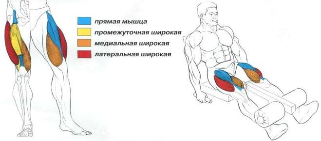
The quadriceps muscle of the thigh (m. Quadriceps femoris) (Fig. 131) consists of four heads and is the largest muscle of a man. With the reduction of all heads, it extends the lower leg, while reducing the rectus femoris muscle, it participates in its flexion. Located on the anterolateral surface of the thigh, in the lower sections completely goes to the side. Each head has its own starting point. The longest rectus femoris muscle (m. Rectus femoris) (Fig. 90, 129, 132, 145) begins on the inferior iliac spine; the medial broad muscle of the thigh (m. vastus medialis) (Fig. 90, 129, 130, 132, 133, 145) - on the medial lip of the rough line of the femur; lateral broad muscle of the femur (m. vastus lateralis) (Fig. 90, 129, 130, 131, 133, 145) - on the greater trochanter, the intertrochanteric line and the lateral lip of the rough line of the femur; the intermediate broad muscle of the femur (m. vastus intermedius) (Fig. 130, 145) - on the anterior surface of the femur. All heads grow together, forming a common tendon, which is attached to the top and side edges of the kneecap, bypassing which the tendon falls below and goes into the knee ligament, which is attached to the tibial tuberosity. In the place of attachment of the muscles, there are the patellar pouch (bursa suprapatellaris), the subcutaneous podrenal bag (bursa subcutanea prepatellaris), the subcutaneous podnatserenny bag (bursa subcutanea infrapatellaris) and the deep undergrown bag (bursa infrapatellar profunda).
The articular muscle of the knee (m. Articularis genus) (Fig. 136) tightens the bag of the knee joint. It is a flat plate and is located on the front surface of the thigh under the intermediate broad muscle of the thigh. The point of its beginning is on the front surface of the lower third of the femur, and the attachment point is on the front and side surfaces of the articular bag of the knee joint.
Medial group
The crest muscle (m. Pectineus) (fig. 90, 129, 130, 132) flexes and leads the thigh, rotating it outwards. The flat muscle is quadrangular in shape, starts on the ridge and upper branch of the pubic bone, and is attached on the medial lip of the rough line of the femur below the small skewer.
Thin muscle (m. Gracilis) (Fig. 90, 129, 130, 132, 134, 145) leads the thigh and takes part in flexing the tibia, turning the leg inward. The long flat muscle is located directly under the skin. The point of its beginning is on the lower branch of the pubic bone, and the place of attachment is on the tibial tuberosity. The tendon of the thin muscle grows together with the tendons of the tailoring and semi-tendinous muscles and the fascia of the tibia, forming a superficial goose foot. Here is the so-called goose bag (bursa anserina).
The long adductor muscle (m. Adductor longus) (Fig. 90, 129, 130, 132) leads the thigh, takes part in its flexion and rotation outwards. This is a flat muscle, having the shape of an irregular triangle and located on the anteromedial surface of the thigh. It starts from the upper branch of the pubic bone and is attached on the middle third of the medial lip of the rough line of the femur.
The short adductor muscle (m. Adductor brevis) (Fig. 131) causes the thigh to participate in its flexion and rotation outwards. It is a muscle of a triangular shape, starts on the front surface of the lower branch of the pubic bone, lateral to the thin muscle, and is attached on the upper third of the medial lip of the rough line of the femur.
The large adductor muscle (m. Adductor magnus) (Fig. 129, 130, 131, 132, 134) leads the thigh, partly rotating it outwards. Thick, wide, the most powerful of this group of muscles, which is located deeper than the rest of the adductor muscles. The point of its beginning is on the sciatic hill, as well as on the branch of the sciatic bone and the lower branch of the pubic bone. The attachment site is located on the medial lip of a rough line and medial epicondyle of the femur. In the muscle bundles, several holes are formed that allow blood vessels to pass through. The largest of these is called the tendon orifice (hiatus tendineus). Above it is a fascial plate, and between it and the muscle a space of triangular shape is formed, called the lead channel (canalis adductorius) (Fig. 131). The femoral vein, artery and hidden nerve of the lower extremity pass through it.
Back group
The biceps muscle of the thigh (m. Biceps femoris) (Fig. 133, 134, 145) extends the thigh and bends the lower leg. In the bent position rotates the shin outwards. Passes along the side edge of the upper surface of the thigh. The muscle has one abdomen and two heads. The long head (caput longum) starts from the sciatic hill, the short head (caput breve) - on the lower part of the lateral lip of the rough line of the femur. The abdomen ends with a long narrow tendon, the attachment of which is located on the head of the fibula. Part of the bundles are woven into the fascia of the leg. Near the point of the beginning of the long muscle is the upper bag of the biceps femoris (bursa m. Bicipitis femoris superior). In the area of the tendon is the lower tummymine sac of the biceps femoris (bursa subtendinea m. Bicipitis femoris inferior).
The semi-tendinous muscle (m. Semitendinosus) (fig. 130, 132, 134, 145) extends the thigh, bends the lower leg, rotates it in a bent position, and also participates in the extension of the body. The muscle is long and thin, partially covered by the gluteus maximus, sometimes interrupted by a tendon bridge (intersectio tendinea) (Fig. 134). The point of its beginning is located on the sciatic tubercle, and the place of attachment is on the medial surface of the tibial tuberosity. Separate bundles of muscles are woven into the fascia of the leg, taking part in the formation of a goose foot.
The semi-membranous muscle (m. Semimembranosus) (fig. 130, 132, 134, 145) extends the thigh and bends the lower leg, rotating it inwards. It passes along the medial edge of the posterior surface of the thigh and is partially covered by the semitendinosus muscle. The muscle starts from the sciatic hill and is attached at the edge of the medial condyle of the tibia.
The tendon is divided into three bundles, forming a deep goose foot. The external bundle passes into the popliteal fascia, into the posterior ligament of the knee joint.
In the place of the tendon division into separate bundles is located the synovial pouch of the semimembranous muscle (bursa m. Semimembranosi).
Atlas of Human Anatomy. Encyclopedias & Dictionaries. 2011 .
Page 3 of 4
MUSCLES OF THE FREE PART OF THE LOWER LIMB
Thigh muscles
The muscles of the thigh are divided into 3 groups: front (hip flexors), back (hip extensors) and medial (resulting thigh).
Having a large mass and a considerable extent, these muscles are able to develop great strength, acting on both hip and knee joints. The thigh muscles perform static and dynamic functions when standing, walking. Like the muscles of the pelvis, the muscles of the thigh reach maximum development in humans due to erect position.
Anterior thigh muscle group
Sartorius (m.sartorius) begins on the superior anterior iliac spine. The muscle crosses obliquely from top to bottom and medially to the front of the thigh. It is attached, passing into the tendon strain, to the tibial tuberosity and to the fascia of the tibia.
At the site of attachment, the tendon of the tailor's muscle coalesces with the tendons of the thin and semi-tendinous muscles and forms a triangular fibrous plate, the so-called goose foot (pes ansermus superficialis), under which there is goose foot bag (bursa anserina).
Function: flexes the thigh and shin, is also involved in turning the hips outwards.
Innervation:
Blood supply: muscle branches of the femoral artery, the lateral artery, the circumflex femur, the descending knee artery.
Fig. 185. Deep thigh muscles; view from the anterior medial side.
1 - ileal muscle (cut off); 2 - large lumbar muscle (cut off); 3 - ilio-comb synovial sac; 4 - comb muscle; 5 - long adductor muscle; 6 - large adductor muscle; 7 - femoral artery and vein; 8 - thin muscle; 9 - tailor muscle (cut off); 10 - the medial broad muscle of the thigh; 11 - rectus femoris; 12 - the gluteus medius muscle (cut off).
Thigh quadriceps muscle (m.quadriceps femoris) strong, has the greatest mass among all muscles.
It consists of 4 muscles forming its head: straight, lateral, medial and intermediate wide muscles of the thigh, which are adjacent to the femur from almost all sides (Fig. 185). In the distal third of the femur, all 4 heads form a common tendon that attaches to the tibial tuberosity, as well as to the top and side edges of the patella. Distally from the top of the patella, the middle part of the tendon continues in patella band (lig. patellae).
Straight muscle of thigh
(m.rectus femoris) begins on the lower anterior iliac spine and on the ilium above the acetabulum. There is a synovial sac between the bone and the beginning of the muscle. Next, the muscle goes down in front of the hip joint, goes to the thigh surface between the muscle - the fastener of the wide fascia and the sartorius muscle, located in front of the intermediate wide thigh muscle. The rectus muscle ends with a tendon, which when
attached to the base of the patella. The muscle has a feathery structure.
Lateral hip muscle (m.vastus lateralis) is the largest of all 4 heads of the quadriceps. It begins with tendon and muscle bundles on the intertrochanteric line, the lower part of the greater trochanter, on the gluteal tuberosity and the upper half of the rough thigh line, and also on the lateral intermuscular hip partition. Attached to the tendon of the rectus femoris, the upper lateral part of the patella and to the tibial tuberosity. Part of the tendon bundles continues in patella lateral ligament support (retinaacum patellae laterаle).
Medial thigh muscle (m.vastus medialis) has an extensive beginning on the lower half of the intertrochanteric line, on the medial lip of the rough line and medial intermuscular partition of the thigh. Attached to the upper edge of the base of the patella and to the anterior surface of the medial condyle of the tibia. The tendon of this muscle is involved in the formation of medial patellar patellar ligament (retinaculum patellae medial).
Intermediate wide hip muscle (m.vastus intermedius) begins with muscle bundles along the upper two-thirds of the anterior and lateral surfaces of the body of the femur, on the lower part of the lateral lip of a rough femur and the lateral intermuscular septum. It is attached to the base of the patella and, together with the tendons of the direct, lateral and medial broad muscles of the thigh, is involved in the formation of the common tendon of the quadriceps femoris.
Function: the quadriceps muscle of the thigh is a powerful extensor of the leg in the knee joint; rectus flexes the thigh.
Innervation: femoral nerve (L II -L IV).
Blood supply: femoral artery, deep femoral artery.
Rear thigh muscle group
The biceps of the thigh, the semitendinosus, and the half-membranous muscles belong to the muscles of the posterior group (see Fig. 182, 183). Proximally at the site of the beginning on the sciatic tubercle, they are covered with a large gluteus muscle. Below, in the posterior region of the femur, the semitendinosity and semimembranosus muscles are located medially, adjacent to the large adductor muscle. The biceps muscle of the thigh occupies the lateral position and is adjacent to the lateral broad muscle of the thigh. Starting from the level of the border between the middle and lower thirds of the thigh, the muscles diverge to the sides, so the semitendinosus and semi-membranous muscles limit the popliteal fossa from the medial side, and the biceps of the thigh - from the lateral one.
Thigh biceps (m.biceps femoris) has two heads - long and short. Long head (cаput longum) together with the semitendinosus muscle begins on the upper medial surface of the sciatic tuber and on the sacro-cuspid ligament, where there is upper bag biceps femur (bursa musculi bicipitis femoris superior). At the level of the lower third of the thigh, the long head of the biceps of the thigh is separated from the semitendinosus muscle and is connected to the short head, passing into a flat tendon. Short head (caput breve) begins on the lateral lip of the rough line, the upper part of the lateral over the condyle and on the lateral intermuscular partition of the thigh. The common muscle tendon is directed down the posterior-lateral side of the knee joint and is attached to the head of the fibula and the outer surface of the lateral condyle of the tibial bone. Part of the tendon bundles continues into the fascia of the leg. Between the tendon of the muscle and the fibular collateral ligament there is inferior shoulder bag of biceps femoris (bursa subtendmea m.bicipitis femoris inferior).
Function: together with other muscles of the back of the group extends the thigh; flexes the lower leg at the knee joint; when bent at the knee joint, the tibia turns it outwards.
Innervation: the long head is the tibial nerve (S I –S II), the short head is the common peroneal nerve (L IV –S I).
Blood supply: the medial artery, the envelope of the femur, the piercing arteries.
Semitendinosus muscle (m.semitendinosus) begins with the long head of the biceps femoris on the ischial tubercle. At the level of the middle third of the thigh goes into the long tendon, which follows down on the posterior-medial side of the knee joint and attaches to the medial surface of the upper part of the tibia (participates in the formation of the superficial goose foot).
Function: extends the thigh, flexes the lower leg; when bent at the knee joint, the shank turns it inwards.
Innervation:
Blood supply: piercing arteries.
Poluponevchataya muscle(m.semimembranosus) begins on the sciatic tubercle with a flat long tendon. The tendon plate continues downward and, narrowing in the distal direction, passes at the level of the mid-thigh into the muscular belly. This abdomen is located anterior to the semitendinosus muscle and the long head of the biceps femoris. At the level of the knee joint, the muscular abdomen continues again into a flat tendon, which is attached with 3 beams to the posterolateral surface of the medial condyle of the tibial bone. These tendon bundles of the semimembranous muscle form the so-called deep goose foot (fig. 186). One tendon bundle continues downward and joins the tibial collateral ligament. The second bundle, following downward and laterally, is interwoven into the fascia of the popliteal muscle, and also attached to the line of soleus muscle of the tibia. The third, the largest tuft, is directed upwards and laterally to the posterior surface of the lateral condyle of the femur, forming an oblique popliteal ligament. Where the tendon of the semimembranous muscle spreads through the medial condyle of the femur and contacts the medial head of the gastrocnemius muscle, there is a synovial bag of this muscle (bursa musculi semimembranosi).
Function: extends the thigh and flexes the shin; when the shank is bent at the knee, it turns it inwards; delays the capsule of the knee joint, protecting the synovial membrane from pinching.
Innervation: tibial nerve (L IV -S I).
Blood supply: artery around the femur, the piercing and popliteal arteries.
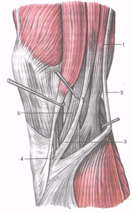
Fig. 186. Tendons of the tailor, thin, semi-contracting and semi-membranous muscles; view from the medial side.
1 - semimembranosus muscle; 2 - tendon of the semitendinosus muscle; 3 - thin muscle tendon; 4 - tailor muscle tendon; 5 - tibial collateral ligament.
Medial thigh muscle group
The muscles of the medial group are thin, comb and leading muscles (long, short and large). The main function of the muscles of this group is the hip reduction, so they are called adductor muscles. They achieve a strong development in humans due to upright walking. These muscles begin on the outer surface of the sciatic and pubic bones, near the obturator foramen. Places of the beginning of the muscles occupy a relatively large surface - from the level of the pubic tubercle to the ischial tubercle.
Leading muscles are attached in the area from the small skewer to the medial epicondyle of the thigh. The general direction of the muscle bundles is oblique; they extend from front to back, from top to bottom, to the rough thigh line, which serves as an attachment site for most of these muscles (Fig. 187).
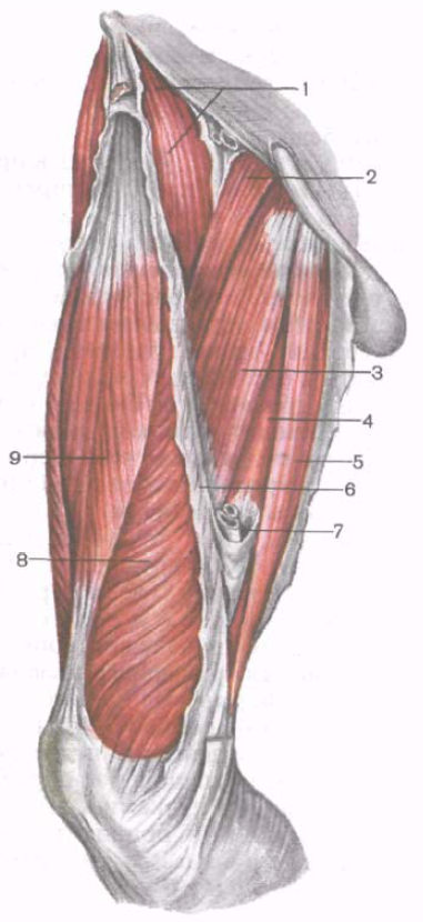
Fig. 187. Muscles of the thigh; anterior and medial groups.
1 - iliopsoas muscle; 2 - comb muscle; 3 - long adductor muscle; 4 - large adductor muscle; 5 - thin muscle; 6 - medial intermuscular partition of the thigh; 7 - the entrance to the leading channel; 8 - medial wide muscle (thighs); 9 - rectus femoris.
Thin muscle (m.grаcilis) flat, long, located superficially along the medial surface of the thigh. It starts with a short tendon on the lower half of the pubic symphysis and on the lower branch of the pubic bone. In the lower third of the thigh, the abdomen is located between the tailor's and semi-membranous muscles. The tendon of the small muscle is attached to the medial surface of the upper body of the tibia and is involved in the formation of the superficial goose foot.
Function: leads the thigh; bends the calf while turning it inwards.
Innervation:
Blood supply: obturator, femoral, and external genital arteries.
Comb muscle (m.pectmeus) short, flat, begins on the crest and upper branch of the pubic bone. Attached with a flat thin tendon to the area located between the back surface of the lesser skewer and the rough thigh line.
Function: participates in adduction and flexion of the hip.
Innervation: obturator nerve (L II -L III).
Blood supply:
Long adductor muscle (m.adductor longus) has a triangular shape, is located medially and downwards from the comb muscle, covers the front adductor muscle and the upper bundles of the major adductor muscle in front. It starts with a thick tendon on the outer surface of the pubic bone (between the crest and the pubic symphysis). Goes down and laterally, goes into a thin wide tendon, which is attached to the medial lip of the rough line of the thigh between the attachment points of the large adductor and the medial broad muscle of the thigh.
Function: leads the thigh, simultaneously bends and turns it outwards.
Innervation: obturator nerve (L II -L III).
Blood supply: obturator and external genital arteries, deep femoral artery.
Short adductor muscle (m.adductor brevis) thick, triangular. It begins on the outer surface of the body and the lower branch of the pubic bone. It is located behind the comb muscle and the long adductor muscle. Heading down and laterally, the muscle expands and attaches with short tendon bundles to the upper part of the rough line.
Function: leads the thigh, is involved in the flexion of the thigh.
Innervation: obturator nerve (L II -L III).
Blood supply: obturator and piercing arteries.
Big adductor muscle (m.adductor mаgnus) thick, triangular shape. It begins on the sciatic tubercle, the branch of the sciatic bone and on the lower branch of the pubic bone. Attached all over the medial lip of the rough line. Located behind the short and long conductive muscles. Behind it are adjacent the semi-tumbler, semi-membranous muscles and the long head of the biceps of the thigh. The proximal muscle tufts are oriented almost horizontally, passing from the pubic bone to the upper part of the thigh. The tufts of the distal part of the muscle are directed vertically downwards - from the ischial tuber to the medial epicondyle of the thigh. The tendon of the major adductor muscle at the site of attachment to resulting tubercle (tuberculum adductorium) of the femur borders a hole called tendon gap(hiatus tendmeus adductorius). Through this gap the femoral artery from the adductor canal on the thigh passes into the popliteal fossa. Next to the artery lies the femoral vein.
Function: is the strongest adductor of the thigh; medial muscle bundles, originating on the sciatic tubercle, are also involved in the extension of the thigh.
Innervation: obturator (L II -L III) and sciatic (L IV -L V) nerves.
Blood supply: obturator and piercing arteries.
Muscles of the lower leg
The muscles of the leg, like other muscles of the lower limb, are well developed, which is determined by the function they perform in connection with erect, static and dynamic of the human body. Having an extensive start on the bones, intermuscular septa and fascia, the calf muscles act on the knee, ankle and foot joints.
There are anterior, posterior and lateral groups of the lower leg muscles. The anterior tibial muscle, the long extensor of the fingers, the long extensor of the thumb belong to the anterior group. The back group includes the triceps muscle of the calf (consisting of the gastrocnemius and soleus muscles), the plantar and popliteal muscles, the long flexor of the fingers, the long flexor of the big toe, the posterior tibialis muscle. The lateral group of the lower leg includes the short and long peroneal muscles.
Anterior leg muscle group
Anterior tibial muscle (m.tibialia anterior) is located on the front side of the tibia. It starts on the lateral condyle and the upper half of the lateral surface of the body of the tibia, as well as the adjacent part of the interosseous membrane and on the fascia of the tibia. At the level of the distal third of the leg, the muscle bundles pass into the long tendon, which passes under the upper and lower retainers of the extensor tendons, anterior to the ankle joint. Next, the tendon goes around the medial edge of the foot and attaches to the plantar surface of the medial cuneiform bone and the base of the first metatarsal bone.
Function: extends the foot in the ankle joint, simultaneously raises the medial edge of the foot and turns it outwards (supination), strengthens the longitudinal arch of the foot. With a fixed foot, bends forward shin; contributes to keeping the tibia upright.
Innervation:
Blood supply; anterior tibial artery.
Fig. 188. Deep muscles of the anterior region of the leg and back of the foot, right.
1 - patella; 2 - interosseous membrane of the leg; 3 - tibia; 4 - muscle - long extensor of the big toe; 5 - tendon of the anterior tibial muscle; 6 - upper extensor muscle tendon retainer; 7 - lower extensor muscle tendon retainer; 8 - muscle - short extensor of the big toe; 9 - dorsal interosseous muscle; 10 - muscle - short extensor fingers; 11 - muscle - long extensor fingers.
Long finger extensor (m.extensor digitorum longus) - pinna muscle, begins on the lateral condyle of the tibia, the front surface of the body of the fibula, on the upper third of the interosseous membrane, fascia and the anterior intermuscular partition of the tibia (Fig. 188). Heading to the rear of the foot, the muscle passes behind the upper and lower extensor tendon retainers. At the level of the ankle joint, the muscle is divided into 4 tendons, which are enclosed in the synovial vagina common to them. Each tendon is attached to the back of the base of the middle and distal phalanges of II-V fingers.
From the lower part of the muscle, a small bundle, called the third fibular muscle (m.peroneus tertius), is separated, the tendon of which is attached to the base of the V metatarsal bone.
Function: extends the II-V fingers in the metatarsophalangeal joints, as well as the foot in the ankle joint. The third fibular muscle raises the lateral edge of the foot. With a reinforced foot, the long extensor of the fingers holds the shin in an upright position.
Innervation: deep fibular nerve (L IV -S I).
Blood supply: anterior tibial artery.
Long extensor of the big toe (m.extensor hallucis longus) is located laterally between the anterior tibial muscle medially and the long extensor of the fingers; partially covered by them in front. It begins on the middle third of the anterior surface of the fibula, interosseous membrane of the tibia. The tendon of the muscle passes down to the rear of the foot under the upper and lower retainers of the extensor tendons in a separate synovial vagina and is attached to the distal phalanx of the big toe. Separate tendon bundles can also be attached to the proximal phalanx.
Function: unbends the big toe; also participates in the extension of the foot in the ankle joint.
Innervation: deep fibular nerve (L IV -S I).
Blood supply: anterior tibial artery.
Back muscle group
The muscles of the back group form two layers - superficial and deep. The superficially lying triceps muscle of the lower leg, which creates the characteristic roundness of the lower leg (Fig. 189), is more strongly developed. The deep layer is formed by a small popliteal muscle and 3 long muscles: a long flexor of the fingers (located most medially), a posterior tibial muscle (occupies an intermediate position) and a long flexor of the big toe (located laterally) (Fig. 190).
The surface layer of the posterior group of the leg muscles
Triceps muscle (m.triceps surae) consists of two muscles - the gastrocnemius muscle, which is located superficially, and the soleus muscle, hidden under the gastrocnemius. The gastrocnemius muscle belongs to the articular muscles, it acts on two joints - the knee and the ankle, while the soleus muscle is monoarticular - acts only on the ankle joint.
Calf muscle (m.gastrocnemius) has two heads: the medial and lateral, the surface layers of which are represented by strong tendon bundles. Lateral head (caput laterale) begins on the outer surface of the lower epiphysis of the femur above the lateral condyle. Medial head (сuput medial) begins on the medial condyle of the femur. Under each head of the gastrocnemius muscle is a synovial sac. Between the lateral head and the capsule of the knee joint is located lateral shrinkage bag gastrocnemius muscle (bursa subtendinea musculi gastrocnemii lateris). Between the medial head and the joint capsule is medial sweetening calf muscle bag (bursa subtendinea musculi gastrocnemii medialis). Both bags, as a rule, communicate with the cavity of the knee joint.
In the middle of the calf, both calf muscles pass into a thick tendon, which narrows down and merges with the soleus muscle tendon, forming calcaneal (achilles) tendon (tendo calcaneus, s. achili), which is attached to the heel bump. Between the tendon and the calcaneus is available bag heel (achilles) tendon (bursa tendinis calcаnei, s.Achillis).
Soleus muscle (m.soleus) is thick, flat, lying under the gastrocnemius muscle. In front of her are the muscles of the deep layer. The soleus muscle has an extensive beginning on the posterior surface of the tibia (on the line of soleus muscle) and on tendon arch (arcus tendineus musculi solei), spreading between the tibial and fibula bones. The soleus muscle has a feathery structure, passes into a flat tendon, which is involved in the formation of the heel tendon.
Function: triceps muscle flexes the calf and foot (plantar flexion); with a fixed foot, holds the shin on the talus, preventing it from tipping over.
Innervation:
Blood supply:
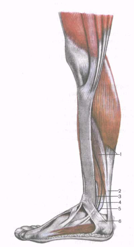
Fig. 189. Muscles of the posterior region of the leg, right; view from the medial side.
1 - triceps muscle of the leg; 2 - heel (Achilles) tendon; 3 - posterior tibial muscle; 4 - muscle - long flexor of the fingers; 5 - muscle - long flexor of the big toe; 6 - flexor tendon retainer.
Fig. 190. Muscles of the posterior region of the leg, right; deep layer.
1 - popliteal muscle; 2 - posterior tibial muscle; 3 - short fibular muscle; 4 - heel (Achilles) tendon (cut off); 5 - muscle - long flexor of the big toe; 6 - muscle - long flexor of the fingers: 7 - soleus muscle (cut off).
Plantar muscle (m.plantаris) is inconstant, has a small abdomen and a long thin tendon. It starts on the lateral epicondyle of the thigh and on the oblique popliteal ligament. The tendon of this muscle passes between the gastrocnemius and soleus muscles, adjacent to the medial edge of the calcaneal tendon, with which it is attached to the calcaneal tubercle.
Function: tightens the capsule of the knee joint, participates in the flexion of the leg and foot.
Innervation: tibial nerve (L IV -S II).
Blood supply: popliteal artery.
Deep layer of the posterior leg muscle group
The deep layer is formed by 4 muscles: the popliteal, long flexor of the fingers, the long flexor of the big toe, and the posterior tibial muscle, which are separated from the soleus muscle by a deep fasciae plate.
Popliteal muscle (m.popliteus) lies deep in the popliteal fossa. It starts with a thick tendon on the outer surface of the lateral condyle of the femur (below the attachment of the fibular collateral ligament). The muscle is adjacent to the posterior surface of the joint capsule and is located below the arcuate popliteal ligament, on which its medial bundles begin. The muscle attaches to a triangular pad on the posterior surface of the tibia, above the line of soleus muscle.
Function: bends the calf, turning it inwards; tightens the capsule of the knee joint, protecting the synovial membrane from pinching.
Innervation: tibial nerve (L IV -S II).
Blood supply: popliteal artery.
Long finger flexor (m.flexor digitorum longus) has a bisexual structure, begins with fleshy tufts on the posterior surface of the body of the tibia below the soleus muscle, as well as on the fascia and the posterior intermuscular partition of the tibia. It is located behind and medial to the posterior tibial muscle. The tendon of the long flexor of the fingers is directed downwards, crossing the tendon of the posterior tibial muscle from behind and from the lateral side. Next, the tendon of the muscle passes to the sole of the foot behind the medial ankle under the flexor tendon retractor in a separate synovial vagina (between the tendons of the posterior tibial muscle medially and the long flexor of the thumb lateral). Then the tendon bends around the back and bottom of the support of the talus. Being located above the short flexor of the fingers, it is divided into 4 separate tendons, which are attached to the distal phalanxes of the II-V fingers, previously perforated the tendons of the short flexor of the fingers (like the tendons of the deep flexor of the fingers on the hand).
Function: bends distal phalanxes of II-V fingers; bends the foot, turning it outwards.
Innervation: tibial nerve (L IV -S II).
Blood supply: posterior tibial artery.
Long flexor of the big toe (m.flexor halucus longus) is a bisected muscle, begins on the lower two-thirds of the body of the fibula, the interosseous membrane, and the posterior intermuscular partition of the tibia. It is located laterally and behind the posterior tibial muscle. The tendon of the long flexor of the big toe passes under the flexor tendon retainer behind the medial malleolus and laterally to the tendon of the long flexor of the fingers in a separate synovial vagina. Next, the tendon of the long flexor of the big toe falls into the same groove on the posterior process of the talus, passing forward under the support of the talus. Reaching the plantar surface of the big toe, the tendon of the long flexor of the thumb attaches to its distal phalanx. On its way to the foot, this tendon intersects with the tendon of the long flexor of the fingers (lies under it). All along the plantar surface of the first metatarsal bone, the tendon of the long flexor of the big toe lies between the medial and lateral abdomens of the short flexor of the big toe.
Function: flexes the big toe, is involved in flexion (supination) and bringing the foot; strengthens the longitudinal arch of the foot.
Innervation: tibial nerve (L IV - S II).
Blood supply: posterior tibial and peroneal arteries.
Rear tibial muscle (m.tibialis posterior) is located deep on the back of the shin between the long flexor of the fingers (medially) and the long flexor of the big toe (laterally). It starts on the posterior surface of the body of the fibula (between the medial crest and the interosseous edge), the lower surface of the lateral condyle and on the upper two thirds of the body of the tibial bone (below the line of soleus muscle) and the interosseous membrane of the tibia.
The muscle continues into a strong tendon, which lies in the groove on the posterior surface of the medial malleolus in front of the tendon of the long flexor of the fingers (under the retainer of the flexor tendons). Turning to the plantar surface of the foot, the tendon attaches to the tuberosity of the scaphoid bone, to all 3 sphenoid bones, as well as to the base of the IV (and sometimes V) of the metatarsal bone.
Function: bends the foot (plantar flexion), causes the foot and supiniruet it.
Innervation: tibial nerve (L IV - S II).
Blood supply: posterior tibial artery.
Lateral group of leg muscles
The lateral group is represented by the long and short peroneal muscles, which are located on the lateral surface of the tibia under the fascia between the anterior and posterior intermuscular septa.
Long fibular muscle (m.peroneus longus) is bony-circular, lies superficially, begins at the head and upper two-thirds of the lateral surface of the fibula, on the lateral condyle of the tibia, fascia of the tibia and on the intermuscular partitions of the tibia. At the level of the ankle joint, the tendon of the muscle, bending around the lateral ankle at the back, passes first under the upper retainer of the tendons of the peroneal muscles in the common synovial vagina with the tendon of the short peroneal muscle, and then in the groove on the heel bone (under the lower retainer of the short fibular muscle, and then in the fissure on the heel bone (under the lower retainer) of the short fibular muscle, and then in the groove on the heel bone (under the retainer) On the sole, the tendon of the long peroneal muscle passes obliquely forward and medially, and lies in the same groove of the cuboid bone in a separate (own) synovial vagina. The tendon attaches to the base of the I and II metatarsal bones and to the medial sphenoid bone.
In those points where the tendon changes its direction (behind the lateral ankle and on the cuboid bone), it usually thickens due to the fibrous cartilage or sesamoid bone formed in its thickness.
Function: bends the foot, lifts its lateral edge (pronation), strengthens the transverse and longitudinal arches of the foot.
Innervation: superficial peroneal nerve (L IV - S I).
Blood supply: lateral lower knee artery, peroneal artery.
Short Fibial Muscle (m.peroneus brevis) two-pedigreed, begins on the lower two-thirds of the lateral surface of the fibula and on the intermuscular partitions of the tibia. The tendon of the muscle passes on the foot behind the lateral ankle under the tendon retainer of the peroneal muscles, lying in the common synovial vagina along with the tendon of the long peroneal muscle. At the lower edge of this holder, the tendon of the short peroneal muscle turns forward and passes along the outer side of the calcaneus under the peroneal block to the site of attachment on the base of the V metatarsal bone.
Function: raises the lateral edge of the foot; prevents the foot from turning inwards; flexes the foot (plantar flexion).
Innervation: superficial peroneal nerve (L IV -S I).
Blood supply: fibular artery.
Muscles of the foot
Along with the tendons of the leg muscles attached to the bones of the foot, which are part of the anterior, posterior, and lateral groups, the foot has its own (short) muscles. These muscles begin and attach within the skeleton of the foot, have complex anatomical, topographic and functional relationships with the tendons of the muscles of the leg, the attachment points of which are located on the bones of the foot. The muscles of the foot are located on her back and on the sole.
The muscles of the rear foot
The muscles of the rear foot lie under the dorsal fascia of the foot and tendons of the long extensor fingers. These are two muscles - a short extensor of the fingers and a short extensor of the big toe (Fig. 191).
Finger Extender (m.extensor digitorum brevis) is an underdeveloped muscle. It starts on the anterior and lateral surfaces of the calcaneus. The muscle passes along the back surface of the foot obliquely forward and medially. Three tendons of this muscle reach II-IV fingers, join from the lateral side to the tendons of the long extensor of the fingers and together with them attach to the bases of the middle and distal phalanges.
Function: together with the tendons of the long extensor of the fingers is involved in the extension of the toes of the foot.
Innervation: deep fibular nerve (L IV - S I)
Blood supply: lateral tarsus and peroneal arteries.
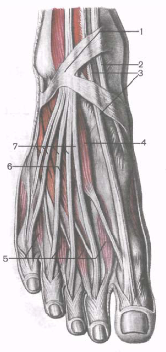
Fig. 191. The tendons of the extensor muscles and short muscles of the rear foot, right.
1 - lower extensor muscles tendon retainer; 2 - tendon of the anterior tibial muscle; 3 - muscle tendon - long extensor of the big toe; 4 -music - short extensor of the big toe; 5 - dorsal interosseous muscles; 6 - muscle - short extensor fingers; 7 - muscle tendons - long extensor fingers.
Short toe extensor (m.extensor hallucis brevis) lies medial to the short extensor of the fingers. It begins on the upper surface of the calcaneus, in its anterior part. The muscle moves forward and medially, passes into the tendon, which is attached to the dorsum of the base of the proximal phalanx of the toe.
Function: involved in the extension of the big toe.
Innervation: deep fibular nerve (L IV -S I).
Blood supply: dorsal artery of the foot.
Muscles of the sole of the foot
In the area of the sole of the foot, the following muscle groups are distinguished: the medial - from the side of the big toe, the lateral - from the little finger, the middle, occupying an intermediate position (Fig. 192, 193).
In contrast to the hand on the sole of the foot, the medial and lateral groups are represented by a smaller number of muscles, and the middle group is strengthened. In general, on the sole 14 short muscles. Three of them belong to the medial group (a muscle that removes the big toe, a short flexor of the big toe, and a muscle that causes the big toe). The two muscles form a lateral group (a muscle that removes the little toe of the foot, and a short flexor of the little toe of the foot). The middle group on the sole reinforced. It consists of 13 muscles. Besides 4 worm-like and 7 interosseous muscles, it includes two more muscles - a short flexor of the fingers and a square muscle of the sole.
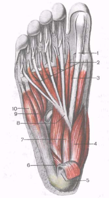
Fig. 192. Muscles of the sole of the foot, right.
1 - muscle tendon - long flexor of the big toe; 2 - worm-like muscles; 3 - muscle - short flexor of the big toe; 4 - muscle, retracting the big toe; 5 - plantar aponeurosis (cut off); 6 - muscle - short flexor of the fingers (cut off); 7 - square muscle of the sole; 8 - muscle tendon - long flexor of the fingers; 9 - muscle - flexor of the little finger of the foot; 10 - the muscle that removes the little finger of the foot.
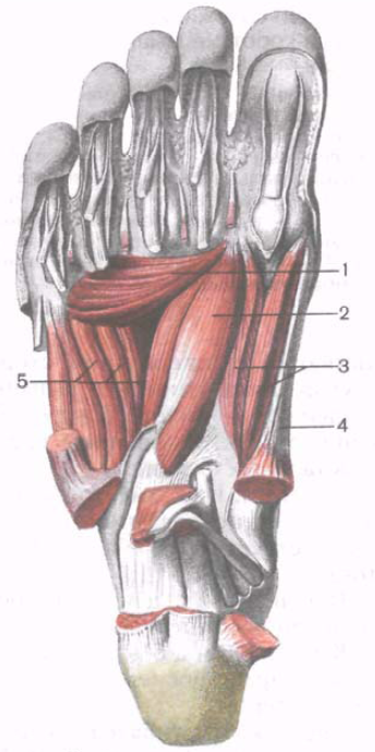
Fig. 193. Deep muscles of the sole of the foot, right. (Superficial muscles and tendons removed.)
1 - muscle leading the big toe (transverse head); 2 - muscle leading the big toe (oblique head); 3 - muscle - short flexor of the big toe; 4 - muscle, retracting the big toe; 5 - plantar interosseous muscles.
Medial group of muscles of the foot
The muscle that removes the big toe (m.abductor halucis), lies superficially along the medial edge of the foot. It begins with short tendon bundles on the medial surface of the calcaneal tuber, and fleshy bundles on the lower retainer of flexor tendons and plantar aponeurosis. The muscle attaches to the medial side of the base of the proximal phalanx of the big toe.
Function: removes the big toe from the midline of the sole of the foot in the medial direction.
Innervation:
Blood supply: medial plantar artery.
Short toe flexor (m.flexor hallucis brevis) adjoins the previous muscle from the lateral side. It starts with a narrow tendinous plate on the medial side of the plantar surface of the cuboid bone (behind the tendon groove of the long peroneal muscle), on the first cuneiform bone and the heeltocube-cuboid ligament. The muscle goes forward and is divided into the medial and lateral parts, between which the tendon of the long flexor of the big toe passes.
Both parts of the muscle are attached to the base of the proximal phalanx and to the sesamoid bones on the sides of the first metatarsophalangeal joint. From the lateral side, the muscle is spliced with the muscle that causes the big toe.
Function: flexes the big toe.
Innervation: the lateral part of the muscle is the lateral plantar nerve (S I -S II); the medial portion is the medial plantar nerve (L V -S I).
Blood supply: medial plantar artery, plantar arch.
The muscle leading the big toe (m.adductor halucis), lies deep, almost in the middle of the sole. It has two heads: oblique and transverse. The oblique head (cataput obliquum) begins on the cuboid, lateral wedge-shaped, on the basis of the II, III and IV metatarsal bones and on the long plantar ligament. The muscular abdomen is directed forward and medially, and passes into a common tendon with the transverse head. Cross head (caput transversum) forms a narrow flat muscular abdomen, which begins on the capsules of the metatarsophalangeal joints of the III-V fingers, goes transversely in the medial direction and connects to the oblique head. The tendon of the adductor is attached to the base of the proximal phalanx of the big toe and to the lateral sesamoid bone.
Function: leads the thumb to the median line of the foot, is involved in bending the big toe of the foot.
Innervation:
Blood supply:
Lateral group of muscles of the foot
The muscle that removes the little toe of the foot (m.abductor digiti minimi), begins with tendon and muscle bundles on the plantar surface of the calcaneus, tuberosity of the V metatarsal bone and on the plantar aponeurosis. The tendon of the muscle passes along the lateral edge of the foot and attaches to the lateral side of the proximal little phalanx.
Function: flexes the proximal phalanx of the little finger and retracts it laterally.
Innervation: lateral plantar nerve (S I -S II).
Blood supply:
Short little foot flexor (m.flexor digiti minimi brevis) begins on the medial side of the plantar surface of the V metatarsal bone, on the vagina of the tendon of the long peroneal muscle, and on the long plantar ligament. The tendon of the muscle lying medially and deeper than the previous one, is attached to the base of the proximal phalanx of the little finger.
Function: bends little finger.
Innervation: lateral plantar nerve (S I -S II).
Blood supply: lateral plantar artery.
Muscle opposing little finger (m.opponens digiti minimi), is located on the lateral side of the short flexor of the little finger. It starts on a long plantar ligament. Attached to the metatarsus V.
Function: involved in strengthening the lateral longitudinal arch of the foot. Muscle is not constant.
Innervation: lateral plantar nerve (S I -S II).
Blood supply: lateral plantar artery.
The middle group of muscles of the foot
Short finger flexor (m.flexor digiti brevis) lies under the plantar aponeurosis. On the lateral side, the muscle is adjacent to the muscle, which removes the little finger, and from the medial side, it is adjacent to the muscle, which removes the big toe. Under the short flexor of the fingers are the square muscle of the sole and the tendon of the long flexor of the fingers. The short flexor of the fingers begins on the front of the plantar surface of the heel bump and on the plantar aponeurosis. From the flat muscular abdomen of this muscle there are 4 tendons that are attached to the middle phalanges of II-V fingers. Each of these tendons at the level of the proximal phalanx splits into two bundles. The long flexor tendon passes through the gap between them. Part of the tendon bundles of the short flexor of the fingers is woven directly into the fibrous sheaths of the toes. The indicated relations of the tendons of the short flexor of the fingers with the tendons of the long flexor of the fingers on the foot are similar to the relations of the tendons of the superficial and deep flexors of the fingers of the hand.
Function: flexes II-V fingers; involved in strengthening the longitudinal arch of the foot.
Innervation: medial plantar nerve (L V -S I).
Blood supply: medial and lateral plantar arteries.
Square sole muscle, extra flexor (m.quadrаtus pаntae, s.m.flexor accessorius) begins on the outer and medial sides of the lower surface of the calcaneus and on the long plantar ligament. The muscle is sent forward and at the level of the middle of the sole of the foot is attached on the lateral side to the tendons of the long flexor of the fingers, heading for the II-IV fingers.
Function: participates in the bending of the toes of the foot, at the same time imparts a straight direction to the long flexor of the fingers.
Innervation: lateral plantar nerve (S I -S II).
Blood supply: lateral plantar artery.
Worm-like muscles (mm.lumbricales); 4 of them have a spindle shape. Laterally lying 3 muscles begin on the facing surfaces of the tendons of the long flexor of the fingers. The fourth, medially located muscle, originates on the medial side of the adjacent tendon of the long flexor of the fingers. Each worm-shaped muscle continues into a thin tendon, which is attached from the medial side to the proximal phalanx of the corresponding finger (II-V). Part of the bundles of tendons of the worm-like muscles bends around the proximal phalanx and passes to the back side of the fingers, interlacing with the tendons of the long extensor of the toes.
Function: bends proximal and unbends middle and distal phalanxes of IV fingers, diverting them medially to the side of the big toe.
Innervation: lateral and medial plantar nerves (L V - S I).
Blood supply: lateral and medial plantar arteries.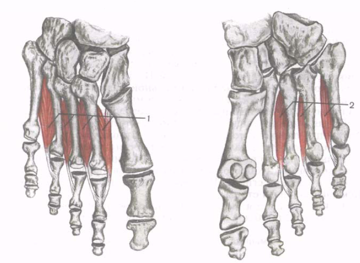
Fig. 194. Interosseous dorsal (1) and plantar (2) muscles.
Intercostal muscles (m.interossei) are located in the intervals between the metatarsal bones (Fig. 194). These muscles are divided into two groups: plantar interosseous and dorsal interosseous muscles.
Unlike similar muscles located on the hand, which are grouped on the sides of the middle finger, on the foot interosseous muscles are concentrated on the sides of the second finger. This is due to the specifics of the function: prehensile - hand and musculoskeletal - feet.
Plantar intercostal muscles (mm.interassei plantаres); there are 3 of them, located in the interosseous spaces on the side of the sole. Each muscle begins on the basis of the medial surface of the bodies of the III-V metatarsal bone. The plantar muscles are attached to the medial surface of the proximal phalanges of the III-V toes of the foot. Part of the bundles passes from the medial side to the dorsal surface of the corresponding finger and weaves into the dorsal aponeurosis.
Function: plantar interosseus muscles lead III-V fingers to finger II; bend the proximal phalanges of these fingers.
Innervation: lateral plantar nerve (S I -S II).
Blood supply: plantar metatarsal arteries, plantar arch.
Rear intercostal muscles (mm.interossei dorsáles); 4 of them, occupy the gaps between the metatarsal bones on the dorsal side. Each dorsal interosseous muscle begins with two heads on the adjacent surfaces of the adjacent metatarsal bones. The tendons of the muscles are attached to the base of the proximal phalanges and to the tendons of the long extensor fingers. The first interosseous muscle is attached to the medial side of the second finger, the other 3 - to the lateral side of the second and fourth fingers.
Function: The first dorsal interosseous muscle retracts the second toe from the midline of the foot to the side of the thumb. The remaining 3 muscles (the second - the fourth) divert II-IV fingers to the lateral side (close to the little finger). The dorsal interosseous muscles flex the proximal phalanges of the II – IV fingers.
Innervation: lateral plantar nerve (S I -S II).
Blood supply: plantar metatarsal arteries, plantar arch.
Questions for repetition and self-control
1. What groups are the muscles of the lower limb girdle? Name the muscles in each such group.
2. What are the muscle groups allocated to the thigh, as well as the muscles in each group?
3. List the synovial bags that are in the area of the knee joint, in the places of the beginning or attachment of muscles.
4. Name the muscles in each of the 3 groups of muscles of the leg.
5. What are the muscles on the rear foot?
6. What muscles are located on the plantar side of the foot? How many of them? Name the function of each of these muscles.
Thanks to the muscles a person has a perfect or not so much silhouette of the legs, pelvis, hips. The brilliant sculptor endowed the thigh muscles with different functions depending on their location. So, the muscles that flex the femur are located on the front part of it - these are the so-called flexors. The muscles that extend the legs are in the posterior segment of the thigh — these are the extensors. The muscle fibers that drive and discharge the hips are located on its inner side and are in the medial group. Consider the anatomy of the femoral muscles and strengthens its exercises.
Anatomy of the thigh muscles
Flexor muscles
The front group joins the muscles in the front segment of the human thigh. This group consists of six muscle fibers: tailor and quadriceps, which include the lateral, straight, intermediate medial, muscles.
Tailor muscle rotates the thigh outward. Tailor fibers are attached to the ilium. In the same place, the tendons of the semitendinosus and the thin muscles grow into a triangle. The tailor's muscle crosses the tibia and ends at the beginning of the tibia. For his development do the following exercises: lunges and squats. At the same time with the tailor, the gluteal muscles of the pelvis are well pumped, especially they can be developed with exercises with a barbell or weights.
The quadriceps muscle is the strongest muscle of the human leg. Quadriceps is formed by four heads. They come from different places of the hip joint and grow into a single tendon of the quadriceps muscle over the patella.
The rectus muscle is its strongest flexor. The rectus muscle of the thigh, which passes right in the middle of the upper leg, grows from the acetabulum and is associated with the tendon below the patella. This is the longest of the 4 quadriceps heads. Its peculiarity is the cirrus structure of the tissue. The rectus muscle of the thigh has short fibers that are stronger than flexible ones. Exercises for this flexor can be performed on a simulator (leg press) and with a barbell (squat).
Medial muscle. This muscle, like most flexors, comes from the femur: from the rough line of its back surface. Medial fibers descend from above into the inside of the human thigh. Exercises that train the medial muscle tissue: squats, better on one leg, lunges, explosive jogging.
The lateral broad muscle of the thigh is responsible for the beautiful shape of the upper legs. This is the largest muscle of the quadriceps. The ideal exercise for their study is the “girlish” squat. With one hand you need to grab hold of something steady, rise on your toes and lower, pushing your knees forward.
The intermediate broad muscle of the thigh was named because of its location: between the medial and lateral muscles. This muscle with the thinnest fibers is under the cover of the rectus femoris muscle. Pumped intermediate press or squats with legs together.
The whole group of quadriceps muscles is strengthened during simple squats. Different position of the feet increases stress.
Muscle extensors
The muscles in the back of the human femoral structures are grouped into the same group. Their composition: biceps femoris muscle (in Latin m.biceps femoris), semi-membranous (m.semimembranosus) and semi-sactimate, (m.semitendinosus). All these muscles begin their growth from the top of the sciatic bone. Combine their purpose and anatomy in a small list.
- The semitendinosus muscle is associated with the tendon with the slender and tailor muscles to the tuber of the tibia. These three muscles form the upper part of the V-shaped fossa under the knee. And the lower segment of the "goose foot" creates the triceps muscle. If the semitendinosus muscle rotates the shin inside, then the three-headed muscle bends the shin at the knee.
- The semimembranosus muscle performs the same action as the semitendinosus. The muscle flexes the inside of the shin, and the hip extends. Half-webbed prevents excessive bending of the knee.
- The biceps muscle of the thigh has two heads. Its fibers grow from two different pelvic bones, from the femoral and sciatic. The biceps muscle of the femur is fastened with a single tendon to the fibula. If you bend the leg at the knee, it will turn the shin out. It is also called the hip biceps.
The muscles of the back surface of the femoral part protect the knee from injuries. But in everyday life, muscles are not sufficiently involved and often tear, because of this, a dent or failure can occur under the skin. Therefore, the semitendinosus, half-membranous and biceps muscular formations of the thigh deserve special attention in training.
Bending, squats with wide feet, benches with legs wide apart, i.e. exercises, where the angle between the body and the femoral part is changed, has a good effect.
Leading muscles
In the medial zone is a muscular group of structures that lead and discharge the human thighs. These muscles are grouped on their inner surface. These include 5 muscles: thin (in Latin m.gracilis), resulting in large (m.adductor magnus), long and short inducing, and also comb. The basic function of such muscles is reflected in the title - hip reduction. Muscles begin to grow from the outer surface of the pelvis, in the area of the pubic tubercle and the obturator opening. Muscle fibers on a rough line of a femur come to an end.
| Muscles | Specifications |
| Thin or slim. | It runs between the tailoring and semi-membranous fibers. This muscle flexes the calf and rotates it inwards. |
| Comb. | It grows from a high point of the pubic bone in the shape of a quadrangle. Its function is to bring and flex the hip joint. If you fix the leg and squeeze the hips, the comb muscle will tilt the pelvis forward. |
| Long leading. | It is located right under the skin. The muscle develops from the lower part of the pubis and ends with the tailoring and semi-tendinous muscles on the tubercle of the tibia bone. The long adductor is shaped like a triangle. The adductor bends and rotates the hip outward. |
| Short lead. | It also has a triangular appearance and runs behind the comb and long adductor muscles. Its fibers reduce the thigh and participate in its rotation. |
| Big lead. | Possesses the most dense fibers. |
![]() The muscle is located deeper than the rest of the adductors, it rotates the thigh outward. A similar function has a square muscle of the thigh, whose fibers are connected with a large resulting under the cover of the gluteus muscle. The square muscle of the thigh comes from the top of the sciatic bone and takes part in the rotation of the thigh.
The muscle is located deeper than the rest of the adductors, it rotates the thigh outward. A similar function has a square muscle of the thigh, whose fibers are connected with a large resulting under the cover of the gluteus muscle. The square muscle of the thigh comes from the top of the sciatic bone and takes part in the rotation of the thigh.
Tailor's muscle (m.sartorius) begins on the superior anterior iliac spine. The muscle crosses obliquely from top to bottom and medially to the front of the thigh. It is attached, passing into the tendon strain, to the tibial tuberosity and to the fascia of the tibia.
The quadriceps muscle (m.quadriceps femoris) is strong, has the greatest mass among all muscles. It consists of 4 muscles forming its head: straight, lateral, medial and intermediate wide muscles of the thigh, which are adjacent to the femur from almost all sides. In the distal third of the thigh, all 4 heads form a common tendon that attaches to the tuberosity of the large tibia, as well as to the top and side edges of the patella. Distally from the top of the patella, the middle part of the tendon continues in patella band(lig. patellae).
Rear thigh muscle group
The biceps of the thigh, the semitendinosus, and the half-membranous muscles belong to the muscles of the posterior group. Proximally at the site of the beginning on the sciatic tubercle, they are covered with a large gluteus muscle. Below, in the posterior region of the femur, the semitendinosity and semimembranosus muscles are located medially, adjacent to the large adductor muscle. The biceps muscle of the thigh occupies the lateral position and is adjacent to the lateral broad muscle of the thigh. Starting from the level of the border between the middle and lower thirds of the thigh, the muscles diverge to the sides, so the semitendinosus and semi-membranous muscles limit the popliteal fossa from the medial side, and the biceps of the thigh - from the lateral one.
The biceps muscle of the thigh (m.biceps femoris) has two heads - long and short. The long head (caput longum), along with the semitendinosus muscle, begins on the upper medial surface of the sciatic tuber and on the sacrospinal ligament, where there is upper bag biceps femur(bursa musculi bicipitis femoris superior). At the level of the lower third of the thigh, the long head of the biceps of the thigh is separated from the semitendinosus muscle and is connected to the short head, passing into a flat tendon.
The semitendinosus (m.semitendinosus) begins with the long head of the biceps femoris on the sciatic tubercle. At the level of the middle third of the thigh goes into the long tendon, which follows down on the posterior-medial side of the knee joint and attaches to the medial surface of the upper part of the tibia (participates in the formation of the superficial goose foot).
The semi-membranous muscle (m.semimembranosus) begins on the sciatic tubercle with a flat long tendon. The tendon plate continues downward and, narrowing in the distal direction, passes at the level of the mid-thigh into the muscular belly. This abdomen is located anterior to the semitendinosus muscle and the long head of the biceps femoris. At the level of the knee joint, the muscular abdomen continues again into a flat tendon, which is attached with 3 beams to the posterolateral surface of the medial condyle of the tibial bone. These tendinous bundles of semimembranous muscle form the so-called deep goose foot.
Medial thigh muscle group
The muscles of the medial group are thin, comb and leading muscles (long, short and large). The main function of the muscles of this group is the hip reduction, so they are called adductor muscles. They achieve a strong development in humans due to upright walking. These muscles begin on the outer surface of the sciatic and pubic bones, near the opening. Places of the beginning of the muscles occupy a relatively large surface - from the level of the pubic tubercle to the ischial tubercle. The adductor muscles are attached in the area from the lesser skewer to the medial sup-condyle of the thigh. The general direction of the muscle bundles is oblique; they extend from front to back, from top to bottom, to the rough thigh line, which serves as an attachment site for most of these muscles.
Thin muscle (m. Gracilis) is flat, long, located superficially along the medial surface of the thigh. It starts with a short tendon on the lower half of the pubic symphysis and on the lower branch of the pubic bone. In the lower third of the thigh, the abdomen is located between the tailor's and semi-membranous muscles. The tendon of the small muscle is attached to the medial surface of the upper body of the tibia and is involved in the formation of the superficial goose foot.
The comb muscle (m.pectineus) is short, flat, begins on the crest and upper branch of the pubic bone. Attached with a flat thin tendon to the area located between the back surface of the lesser skewer and the rough thigh line.
The long adductor muscle (m.adductor longus) has a triangular shape, is located medially and downward from the comb muscle, covers the short adductor muscle and the upper bundles of the large adductor muscle in front. It starts with a thick tendon on the outer surface of the pubic bone (between the crest and the pubic symphysis). The short adductor muscle (m.adductor brevis) is thick, triangular in shape. It begins on the outer surface of the body and the lower branch of the pubic bone. It is located behind the comb muscle and the long adductor muscle. Heading down and laterally, the muscle expands and attaches with short tendon bundles to the upper part of the rough line.
The large adductor muscle (m.adductor magnus) is thick, triangular in shape. It begins on the sciatic tubercle, the branch of the sciatic bone and on the lower branch of the pubic bone. Attached all over the medial lip of the rough line. Located behind the short and long conductive muscles. Behind it are adjacent the semi-tendon, semi-membranous muscles and the long head of the biceps of the thigh. The proximal muscle tufts are oriented almost horizontally, passing from the pubic bone to the upper part of the thigh.
The muscles of the thigh are very well developed in connection with upright walking. They are massive, act on the hip and knee joints, participate in the functions of standing, walking, jogging. These muscles are not only involved in the movement of the body, but also keep it upright. The muscles of the thigh are divided into 3 groups. The anterior group (hip flexors and shin extensors) consists of two muscles — a quadriceps and a tailor; the posterior (extensors of the thigh and flexors of the lower leg) consists of three muscles - a semitendinosus, a half-membranous, and two-headed; The medial group (adductor femur) is formed by five muscles — it is a comb muscle, a thin, long, short, and large adductor muscle. In connection with the erect position, flexion in the knee joint in a person is facilitated, since this is facilitated by the force of gravity. Thigh flexor muscles are less developed, and the quadriceps, which is the extensor of the lower leg, is better developed.
Anterior thigh muscle group. Sartorius(m. sartorius),ribbon-like, up to 50 cm long, located on the front side of the thigh, in the groove between the quadriceps and the adductors of the thigh. Tailor's muscle begins on the upper anterior iliac spine of the Ilium, anterior to the muscle that strains the wide fascia of the thigh. Further tailor muscle obliquely downwards and medially
crosses the front of the thigh, being enclosed in a separate fascial sheath of the wide fascia of the thigh. Just above the knee joint, the muscle passes into a tendon strain, which attaches to the tibial tuberosity and is woven into the fascia of the tibia. At the site of attachment, the tendon of the tailor's muscle coalesces with the tendons of the thin and semi-tendinous muscles. As a result, together with them a triangular plate is formed - goose foot (pes anserinus superficialis),under which is located the same name mucous bag (bursa ansertinae crturis).
Function:flexes the thigh and shin, retracts and rotates the thigh outwards, and when the lower limb is raised and the lower leg bent at the knee, turns the lower leg inward.
Innervation:femoral nerve (LII-LIV).
Blood supply:lateral artery, surrounding femur, descending knee artery.
Thigh quadriceps muscle(m. quadri ceps femoris)the strongest and most powerful of all the muscles, located on the front side of the thigh (see fig. 162, 165). It consists of four parts (muscles): the rectus, lateral, medial and intermediate wide muscles of the thigh, which are almost on all sides adjacent to the femur. In the lower third of the thigh, all four muscles join and form a tendon common to them. This wide and thick tendon attaches to the tibial tuberosity, as well as to the base, top, and side edges of the patella. The tendon between the patella and tibia is called patella ligaments (lig. pattellae).Thus, the patella, located in the thickness of the quadriceps tendon, is a sesamoid bone that increases the attachment angle of this tendon to the tibial tuberosity. Ahead of the patella below the tendon is podsuhozhilnaya pre-knee bag (bursa prepatellaris subtendinea).
Under the tendon of the quadriceps, at the place of attachment to the tibial tuberosity, is deep patellar bag (bursa infrapatellaris profunda).
The tendon fibers of the medial and lateral wide thigh muscles not only join the tendon of the rectus femoris muscle, participating in the formation of the common tendon, but also attach themselves to the edges of the patella, forming medial and lateral tendon strains- medial and lateral support of the patella ligament.
Straight muscle of thigh(m. rectus femoris)- the longest of all parts of the quadriceps muscle of the thigh, spindle-shaped, two-pedigree. Begins
on the lower anterior iliac spine and on the outer surface of the ilium above the acetabulum. Between the beginning of the muscle and the lower front iliac awn is bag straight muscle of the thigh (bursa mtosculi recti femoris).The muscle goes from top to bottom, in front of the hip joint, goes to the front of the thigh, where it passes into the common tendon of the quadriceps muscle of the thigh.
Lateral hip muscle(m. vastus lateralis)- the largest of all four parts of the quadriceps muscle of the thigh, located on its posterolateral side. This muscle is covered by a muscle fastener of the wide fascia of the thigh and the iliac-tibial tract. The lateral broad muscle of the femur is single-pinned, flat, and begins with tendon muscle bundles on the intertrochanteric line of the femur, the lower part of the greater trochanter, gluteal roughness, on the lateral lip of the rough line of the femur and on the lateral intermuscular hip partition. The muscle follows obliquely downwards and medially to the patella and passes into the common tendon. Part of the bundles of this muscle continues in lateral patellar ligament support (retintaculum pattellae latertale).
The medial broad muscle of the thigh (m. Vastus medialis),also single-faced, flat, located on the anteromedial side of thigh. At the top and back of the medial broad muscle of the thigh adjacent to the large and long leading muscles, sometimes even spliced with them. The medial surface of the muscle is covered with tailor muscle. The medial broad muscle of the thigh begins on the lower half of the intertrochanteric line, the medial lip of the rough line of the femur, the medial intermuscular partition of the thigh. Then the muscle follows obliquely down, goes into the common tendon. Part of the tendon bundles involved in the formation of medial patellar ligament (retintaculum pattellae meditale).
Intermediate wide hip muscle (m. Vtastus intermtedius)it is a flat plate, which is spliced laterally and medially with the broad muscles of the thigh, being covered with their edges. In front the intermediate broad muscle of the thigh is covered by the rectus femoris. The intermediate muscle is the weakest of the four muscles that make up the quadriceps muscle of the thigh. It begins with muscle bundles on the upper two thirds of the anterior and lateral surfaces of the body of the femur, the lower part of the lateral lip of the rough line and on the lateral intermuscular partition of the thigh. It should be from top to bottom and goes into the common tendon of the quadriceps femoris.
Part of the deep muscle bundles of the intermediate broad thigh muscle in the lower part of the thigh is attached to the upper and lateral sections.
articular knee pouch. These muscle bundles are called articular muscles of the knee (m. articultaris gtenus).
Function:the quadriceps muscle of the thigh is a powerful extension of the leg in the knee joint (the rectus muscle also flexes the hip). Muscle plays an important role in upright posture and keeping the body in an upright position, counteracting the force of gravity, which tends to bend the leg at the knee joint.
Innervation:femoral nerve (LII-LIV).
Blood supply:femoral artery, deep femoral artery.
Back thigh muscle group. To the posterior thigh muscle group biceps femoris, semiquinatation and semimembranosus,they all have a beginning on the ischial tubercle. At its beginning, these muscles are covered with the gluteus maximus muscle. Semitendinosity and semimembranosus muscles, adjacent behind the large adductor muscle, lie medially. The biceps muscle is lateral. It borders on the lateral broad thigh muscle. On the border between the middle and lower thirds of the thigh muscles diverge. Semitendinosus and semimembranosus muscles go down and medially, limiting the popliteal fossa from the medial side. The biceps muscle of the thigh forms the lateral wall of the popliteal fossa (see. Fig. 163).
Thigh biceps(m. biceps femoris),long, powerful, has two heads: long and short. Long head (caput longum)begins with a thick short tendon (together with the semitendinosus muscle) on the upper medial surface of the sciatic tuber and on the sacrospinal ligament. Then this muscle follows from the top downward obliquely in the lateral direction near the semitendinosus muscle. At the border between the middle and lower thirds of the thigh, the long head of the biceps muscle is separated from the semitendinosus muscle and is connected to its short head. Short head(caput breve)the biceps femoris begins on the lateral lip of the rough line, the upper part of the lateral epicondyle of the femur and on the lateral intermuscular partition of the thigh. Both heads, connecting at the border between the middle and lower parts of the thigh, pass into the common tendon, which follows down the posterolateral side of the knee joint. The tendon is attached to the head of the fibula and to the outer surface of the lateral condyle of the tibia. Some of the fibers of this tendon are woven into the fascia of the leg. Between the biceps tendon and the fibular collateral ligament is inferior lower leg sinew sac of biceps femoris (btorsa subtendinea m. bicipitis femoris inferior). Upper biceps femoris bag (btorsa m.
bicipitis femoris superior)is located between the beginning of the long head of this muscle and the sciatic hill.
Function:straightens and leads the thigh, bends the shin at the knee joint, the shank bent at the knee joint turns outwards.
Innervation:tibial nerve (SI-SII) (long head), common peroneal nerve (LIV-SI) (short head).
Blood supply:the medial artery surrounding the femur; piercing arteries.
Semitendinosus muscle(m. semitendinosus),long, flat, tapering downwards, begins with the long head of the biceps femoris on the upper medial surface of the sciatic tuber and the sacrum-angled ligament. The muscle is located closer to the medial edge of the back of the thigh. The lateral side of the muscle is bordered by the biceps muscle of the thigh, the medial side with the semimembranosus. The proximal part of the semitendinosus muscle is covered with the gluteus maximus muscle. Sometimes, around the middle of the thigh, the semitendinosus muscle has a tendon bridge. The semi-tendinous muscle follows from the top down and at the level of the middle third of the thigh turns into a long tendon. This tendon runs down the posterior-medial side of the knee joint, bends around the back of the femoral epicondyle of the medial fossa and attaches to the medial surface of the upper part of the tibia and fascia of the tibia, participating in the formation of the surface "goose foot".
Function:extends the thigh, the knee bent at the knee turns inwards.
Innervation:tibial nerve (LIV-SII).
Blood supply:piercing arteries.
Poluponevchataya muscle(m. semimembranosus),flat, begins on the sciatic tubercle between the beginning of the semitendinosum and the large adductor muscles with a long flat tendon. The muscle follows from the top down and, narrowing, at the level of the mid-thigh passes into the muscular abdomen, located anterior to the semitendinosus muscle and the long head of the biceps of the thigh. The tendon of the semimembranous muscle, first flattened and then taking a rounded shape, is formed on the medial side of her abdomen. The tendon passes behind the medial epicondyle of the femur. At the level of the posterior surface of the knee joint, the tendon flattens and divides into 3 tendon bundles (medial, middle and lateral), and therefore this branched part of the tendon is called deep goose foot (pes anserinus profundus).The medial (anterior) tendon bundle goes horizontally forward and attaches to the medial condyle
tibia, under the tibial (medial) collateral ligament of the knee joint. The middle tendon bundle is attached to the posterior surface of the medial condyle of the tibia and is woven into the fascia of the popliteal muscle. The lateral (posterior) tendon bundle bends steeply into the lateral side and upwards and continues into the oblique popliteal ligament, which is interwoven into the back wall of the joint capsule of the knee joint.
Under the tendon of the semimembranous muscle in the place of its division into three tendon bundles there is bag of semimembranosus (bursa m. semimembranosi).
Function:extends the hip and bends the shin, bent at the knee joint; the shin turns medially, delays the capsule of the knee joint, and when bent, protects the synovial membrane of the knee joint from infringement.
The muscles of the posterior group play an important role in maintaining the erect position and erect position, preventing the body from bending in the hip joint under the influence of gravity.
Innervation:tibial nerve (LIV-SI).
Blood supply:medial artery, surrounding femur, piercing arteries, popliteal artery.
Medial thigh muscle group. The muscles of the medial group are thin, comb, large, long and short adducting thigh muscles(fig. 165). These muscles, which cause the thigh, are very developed in man in connection with erect walking. The muscles are located on the medial side of the thigh along its entire length. They begin on the bones of the pelvis (the pubic and sciatic) near the obturator orifice, which these muscles cover in front. Under the initial part of the adductor muscles is the external obturator muscle. Comb and thin muscles lie more superficially in the adductor group, the longer adductor muscle lies deeper, and the large and short adductor muscles lie deeper. The muscles of the medial group are attached throughout almost the entire length of the femur, from the lesser trochanter to the medial epicondyle of the thigh.
Thin muscle(m. gracilis),flat, long, ribbon-like, located superficially along the medial side of the thigh. It begins with a short thin tendon on the lower half of the pubic symphysis and on the lower branch of the pubic bone. Next, the muscle should be from top to bottom, located on top near the long adductor muscle (medially) and the large adductor muscle (behind). In the lower third of the thigh, the thin muscle is located between the tailor's anterior muscle and the semimembranosus muscle in the back. At the level of the knee joint thin
Fig. 165.The leading muscle of the thigh, right, view from the medial side:
1 - large adductor muscle; 2 - semitendinosus muscle; 3 - leading channel; 4 - semimembranosus muscle; 5 - medial wide muscle of the thigh; 6 - fibrous plate; 7 - rectus femoris; 8 - long adductor muscle; 9 - short adductor muscle; 10 - comb muscle;
11 - locking membrane;
12 - ileal muscle;
13 - large lumbar muscle; 14 - pear muscle; 15 - the sacral osis
taya ligament
the muscle lies between the tailor's and the semitendinous muscles. The tendon of the small muscle is attached to the medial surface of the upper body of the tibia, participating in the formation of the superficial goose foot along with the tendons of the semitendinosus and sartorius muscles.
Function:leads the thigh, bends the shin at the knee joint, at the same time turning it inwards.
Innervation:obturator nerve (LII-LIV).
Blood supply:obturator, external genital, femoral arteries.
Comb muscle(m. pectineus),short, flat, adjacent to the front surface of the external obturator muscle and to the short adductor muscle. The comb muscle begins on the crest and upper branch of the pubic bone. It should be down and lateral, adjacent to the external obturator muscle and, in part, to the short adductor muscle. The lateral edge of the comb muscle is bordered by the iliopsoas muscle, forming in the place of contact the ilio-comb furrow. The medial edge of the comb muscle is in contact with the long adductor muscle and the hip joint capsule. The flat thin tendon of the comb muscle is attached to the comb line of the femur, located between the posterior surface of the lesser skewer and the rough line of the thigh.
Function:leads and flexes the thigh.
Innervation:
Blood supply:
Long adductor muscle(m. adductor longus),thick, flat, triangular, located medially and downward from the comb muscle, closes the front adductor muscle and the upper bundles of the major adductor muscle in front (see Fig. 165). It begins with a thick, strong tendon on the outer surface of the upper branch of the pubic bone between the pubic tubercle and the pubic symphysis, lateral to the onset of the thin muscle. The long adductor extends from the top down and laterally, continues into the wide flat tendon, which attaches to the middle third of the medial lip of the rough line of the femur, between the attachment zones of the large adductor and medial wide muscles of the thigh.
Function:leads the thigh, simultaneously bends and turns it outwards.
Innervation:obturator nerve (LII-LIII).
Blood supply:obturator, external genital artery, deep femoral artery.
Short adductor muscle(m. adductor brevis),thick, flat, triangular. Located behind the comb and long adductor muscles. It begins on the outer surface of the body and the lower branch of the pubic bone, lateral to the onset of the thin muscle. The muscle goes down and laterally, gradually expands and attaches with short thick tendon bundles to the upper part of the medial lip and the rough line of the femur, behind the attachment point of the long adductor.
Function:causes the thigh, is involved in the flexion and rotation of the thigh outwards.
Innervation:obturator nerve (LII-LIII).
Blood supply:obturator, piercing arteries.
Big adductor muscle(m. adductor magnus),massive, flat, triangular, the largest of all the muscles of the medial group of the thigh. It starts with a short wide tendon on the sciatic tubercle, the branch of the sciatic bone and the lower branch of the pubic bone. The large adductor muscle is located behind the short and long adductor muscles, in front of the lower bundles of the gluteus maximus, the semitendinosus, the semimembranosus, and the long head of the biceps of the thigh. The muscle fan-like expands downwards and medially, its upper beams are directed almost horizontally, following from the pubic bone to the upper part of the body of the femur. Distal muscle bundles are directed downward from the sciatic tuberosum to the medial epicondyle of the femur. The muscle is attached to the medial lip of the rough line of the femur along its entire length, up to its medial epicondyle. At the site of attachment of the tendon bundle of this muscle to the so-called resulting tubercle of the medial condyle of the femur is formed tendon gap,through which the femoral artery passes into the popliteal fossa from the adducting canal on the thigh. Just above this hole is intermuscular tendon plate (lamina vastoadductoria),which in the form of a bridge connects the large adductor muscle and the medial broad muscle, one of the quadriceps muscles of the thigh. The upper bundles of the large afferent muscles, located almost horizontally, are known as small adductor muscle (m. adductor minimus).These muscle bundles are located behind the short adductor muscle, at the top they are adjacent to the external locking muscle and the square muscle of the thigh.