White blood cells
Shear index - The ratio of indicators: (myelocytes + metamyelocytes + band neutrophils) / segmented neutrophils. Normally, the shear index is 0.06.
Increased neutrophil level (neutrophilia, neutrophilia) indicates the presence of an infectious or oncological disease, an inflammatory process, occurs after surgical interventions, with ischemic heart attacks of internal organs (myocardium, kidneys, etc.), endogenous intoxication (uremia), taking a number of drugs (glucocorticoids) , preparations of digitalis, heparin sodium, acetylcholine), poisoning, as well as physical exertion and emotional stress.
Neutropenia (granulocytopenia) - a decrease in the number of neutrophils. Isolated neutropenia, caused by a deficiency of granulocyte progenitors in the bone marrow, may be congenital or acquired.
● Congenital autosomal recessive neutropenia combined with pancreatic insufficiency - Shwachman – Dayemond – Oski syndrome. Repeated infections with steatorrhea in the first years of life are characteristic.
● Acquired absolute granulocytopenia (less 1,8-10 9 / l) is at pertussis, infectious mononucleosis, typhoid, panmielopatii, acute leukemia, severe infectious and toxic processes (sepsis, diphtheria), immune granulocytopenia occurring under the influence antileykotsitarnyh antibodies ( auto- and isoantibodies), after radiation or cytostatic therapy, in the treatment of drugs that are toxic to granulocytopoiesis, the action of benzene, aniline, nitrophenol, etc.
Diagnostic significance of changes in individual hemogram indicators
|
Sign of |
States |
|
Neutrophilic leukocytosis |
Acute infectious and inflammatory diseases, aggravation of chronic diseases, chronic and acute myeloid leukemia, malignant neoplasm of non-hematopoietic organs (carcinoma, sarcoma) in the phase of tumor destruction, erythroleukemia, acute posthemorrhagic anemia, the height of transplant rejection, burns, early period after major surgery, early phase massive radiation damage, coma (uremic, diabetic, hepatic coma), arsenic intoxication, carbon monoxide, epilepsy |
|
Lymphocytic leukocytosis |
Completion of infectious and inflammatory diseases, a number of viral infections (epidemic parotitis, papatachi fever, whooping cough), acute and chronic lymphoblastosis, severe thyrotoxicosis (very rare), chronic radiation sickness |
|
Leukocytosis with absolute osinophilia |
|
|
Leukopenia with absolute neutropenia |
Decompensation of severe infectious and inflammatory processes, rarely remission of chronic inflammatory diseases (tuberculosis, gonorrhea, etc.). Avitaminosis (scurvy, pellagra, etc.). Cachexia, dystrophy, fasting. Cytostatic disease. Chronic benign familial neutropenia. Chronic benign granulocytopenia of childhood (chronic recurrent pediatric agranulocytosis). Cyclic neutropenia, autoimmune leukopenia. Chronic benzene intoxication. Hyperchromic macrocytic anemia (B 12 -deficiency). Hypersplenism. Leukopenic variants of acute leukemia, chronic lymphocytic leukemia |
|
Leukopenia with absolute lymphocytopenia |
Radiation sickness (severe form), cytostatic illness, acquired immunodeficiency syndrome, chronic aleukemic myelosis, leukopenic forms of chronic lymphocytic leukemia |
|
Monocytosis |
Mononucleosis, monocytic leukemia, viral hepatitis, tuberculosis, most autoimmune processes (rheumatic endocarditis) |
|
Monocytopenia |
Severe septic processes, leukemias |
|
Neutrophilis without left shift |
Physiological neutrophilia (physical and emotional stress, food intake, etc.), seizures, epilepsy, weak inflammatory processes (superficial infections, polyarthritis), early stages of uncomplicated tumors, mild thyrotoxicosis |
|
Neutrophilia with a weak or moderate left shift |
|
|
Neutrophilis with a pronounced shift to the left |
Extensive inflammatory processes caused by highly pathogenic microorganisms |
|
Neutropenia |
Severe inflammatory process, autoimmune neutropenia. Neutropenias with a shift of the nuclear formula to the right (overripe hypersegmented forms of neutrophils prevail): B 12 - deficiency anemia, avitaminosis, cachexia, starvation |
|
Eosinophilia |
|
|
Lymphocytopenia |
Leukemia (myeloid leukemia, eosinophilic leukemia). Hereditary eosinophilia. Lymphopenia with relative eosinophilia (pediatric genetic agranulocytosis, B12-deficiency anemia, chronic benign hereditary neutropenia) |
|
Eosinopenia |
The beginning of an infectious disease, intoxication with chemical compounds, heavy metals |
|
Basophilia |
Chronic myeloid leukemia (in parallel with eosinophilia), erythremia, chronic ulcerative colitis, erythroderma, etc. |
The previously indicated changes in the ratio of mature and immature neutrophil forms can be quantified by calculating nuclear shift index. It reflects the ratio of the percentage of the sum of all young forms of neutrophils (stab, metamyelocytes, myelocytes, promyelocytes) to their mature forms.
In healthy adults nuclear shift index ranges from 0.05 to 0.10. Increasing it indicates a nuclear shift of neutrophils to the left, a decrease indicates a shift to the right.
Redistributive and hemoconcentration (false) leukocytosis not accompanied by a change in leukocyte formula.
With significant leukocytosis signs of hyperplasia of lymphopoietic tissue in the form of an increase in the size of lymphoid follicles and their germinal centers are noted in punctates of the bone marrow and lymph nodes.
Types and value of leukocytosis
Types and significance of leukocytosis are shown in the figure.
Physiological leukocytosis.
These include most of the leukocytosis. They are characterized by an adaptive nature and adequacy to the factors causing them. Among physiological leukocytosis, there are functional and protective-adaptive.
- Functional leukocytosis. It is caused by the body performing a certain function (for example, leukocytosis during pregnancy, an increase in the number of leukocytes in the blood of the intestinal vessels after eating or muscles after prolonged physical work).
- Protective and adaptive. It develops during inflammatory processes, damage to cells and tissues (for example, after heart attacks or strokes, soft tissue injuries), stress reaction.
In these and other similar cases, leukocytosis is accompanied by activation of the functions of leukocytes, including one of the most important among them, phagocytic. This helps to increase the body's resistance to infectious and non-infectious pathogenic effects.
Pathological Leukocytosis. Observed with leukemia. This kind of leukocytosis, which develops due to an increase in the number of leukocytes of a tumorous nature, does not have an adaptive value for the organism. Leukemia leukocytes are characterized by impaired functional activity of leukocytes: their ability to synthesize and release cytokines is reduced, and their phagocytic activity is low. In this regard, in patients with leukemia, the effectiveness of immunity reactions is reduced, allergic reactions and diseases of immune auto-aggression are often developed.
Neutrophilia - this is an increase in the content of neutrophils above 6x10 9 / l:
small neutrophilia with a slight shift to the left (with an increase in the percentage of band-leukocytes, sometimes with the advent of metamyelocytes) indicates a mild form of infectious or inflammatory inflammatory disease (usually limited);
significant neutrophilia with a sharp shift to the left (to myelocytes) indicates severe infection (peritonitis, sepsis);
significant neutrophilia with hyperleukocytosis suggests severe septic infection or purulent-inflammatory process with good body resistance;
a sharp neutrophilia with a small leukocytosis indicates severe septic infection with a weakened body resistance;
high neutrophilia in leukopenia is an indicator of severe infection and poor immune resistance.
What are the clinical forms in which neutrophilic leukocytosis is observed?
Acute bacterial infections:
localized (abscesses, osteomyelitis, acute appendicitis, acute otitis, pneumonia (especially lobar), acute pyelonephritis, meningitis, sore throats, acute cholecystitis, etc.);
generalized (septicemia, peritonitis, cholera, etc.).
Inflammation or necrosis of tissue (myocardial infarction, extensive burns, gangrene, malignant tumor with decay, acute attack of rheumatism).
Intoxication:
exogenous (lead, snake venom, vaccines - foreign protein);
endogenous (diabetic acidosis, uremia).
Medicinal effects (corticosteroids, lithium preparations, camphor).
Myeloproliferative diseases (chronic myeloid leukemia, true erythremia, osteomyelofibrosis).
Malignant tumors (stomach, bronchogenic cancer)
Acute hemorrhages.
Diagnostic value of neutrophilic left shift
Neutrophil shift to the left - This is the rejuvenation of the composition of neutrophils, the appearance of immature forms: an increase in the percentage of stab cells, the appearance of metamyelocytes (young), myelocytes, promyelocytes, and sometimes even myeloblasts. Neutrophil shift to the left has several forms. The form of the neutrophilic shift is an important criterion determining the severity of the infection and the prognosis of the disease.
Forms of neutrophilic shift:
leukemoid neutrophilic shift - there is a small percentage of myelocytes, metamyelocytes, sometimes promyelocytes;
regenerative shift - characterized by an increase in the content of stab neutrophils and adolescents;
degenerative - often combined with regenerative, characterized by degenerative changes in the nuclei and cytoplasm of neutrophils. The degree of regenerative shift is determined by the formula:
M + Y + P
Where: M - myelocytes, Yu - young, P - bandwith, With - segmented.
The value of this index is normal 0.05-0.08.
The shift index from 1 to 2 is observed in the presence of very serious infections, purulent-septic diseases;
The shift index from 0.3 to 0.5 is observed in purulent-inflammatory diseases and infections of moderate severity;
A shift index of 0.08 - 0.3 is characteristic of lung diseases.
Diagnostic value of neutrophilic shift to the right
Neutrophil shift to the rightcharacterized by the predominance of mature forms of neutrophils (segmented) with five to six segments. The shift index in this case is 0.04-0.03. In infectious diseases, limited inflammatory processes, the appearance of a neutrophil shift to the right indicates a favorable course of the disease.
Diagnostic value of neutropenia
Neutropenia - this is a decrease in the content of neutrophils less than 2.0 x10 9 / l.
Neutropenia is often a sign of functional inhibition of the bone marrow.
What are the clinical forms in which neutropenia is observed, the mechanisms of its development?
Infections:
bacterial (typhoid fever, paratyphoid fever, tularemia, brucellosis, subacute bacterial endocarditis, miliary tuberculosis);
viral - infectious hepatitis, influenza, measles, rubella.
Toxic effect on the bone marrow with granulocytopoiesis depression:
ionizing radiation;
chemical agents - benzene, aniline, DDT.
Lack of vitamin B 12 and folic acid - essential components of thymidine biosynthesis in bone marrow cells.
Acute aleukemic leukemia, aplastic anemia.
Splenomegaly (enlargement of the spleen of various origins) - due to the "depressive effect" of the spleen on the bone marrow, sequestration and destruction of neutrophils in the spleen and the formation of anti-leukocyte antibodies.
Autoimmune processes - systemic lupus erythematosus, rheumatoid arthritis.
After exposure to certain drugs: analgesics and anti-inflammatory; antiarrhythmic (quinidine, procainamide); antimalarial; anticonvulsants; antihypertensives (captopril, enalapril, representatives of nifedipins); antibacterial (sulfonamides, penicillins, cephalosporins).
Diagnostic value of eosinophilia
Normally, eosinophils constitute 0.5-5% of all leukocytes in peripheral blood (less than 0.4x10 9 / l). Eosinophils live up to 2 weeks. Eosinophils can actively phagocytize. They are actively moving to places of accumulation of antigens, capable of absorbing and transporting them. Eosinophils in their granules contain substances of antihistamine action and this is due to their antitoxic effect during allergic processes.
Eosinophilia called an increase in the content of eosinophils in the blood above 5%. Hyperosinophilia is characterized by numbers in the 20-30% and above. Eosinophilia is observed in allergic processes, worms invasion, some malignant tumors, massive antibacterial therapy.
Name the clinical forms accompanied by eosinophilia, indicating the main causes of its occurrence.
Eosinophiliaobserved under the following conditions:
With allergies (bronchial asthma, hay fever, angioedema, atopic dermatitis, urticaria), intolerance to some drugs (iodine, antibiotics from the penicillin group, etc.).
When worm infestation (trichinosis, echinococcosis, ascariasis, hookworm, opisthorchiasis, wide lentets).
In diseases that occur with the formation of granulomas (lymphogranulomatosis, tuberculosis, syphilis, rheumatism).
For tumors: hemoblastosis (chronic myeloid leukemia, erythremia), for liver cancer, ovarian tumors, uterus.
In some systemic diseases, for example, in systemic lupus erythematosus.
In skin diseases (eczema, psoriasis).
Diagnostic value of basophilia
Basophils normally make up 0-1% of all leukocytes in peripheral blood. Basophilic leukocytes are involved in inflammation and allergic reactions.
Basophilia - this is an increase in the content of basophils in the peripheral blood above 1%. It is observed in chronic myeloid leukemia, erythremia, chronic ulcerative colitis, hypothyroidism, malignant tumors, influenza, chickenpox, and tuberculosis.
Diagnostic value of lymphocytosis
Normally, lymphocytes make up 19-37% of all leukocytes in peripheral blood (1.2-3.0x10 9 / l). 75% of circulating lymphocytes are T-lymphocytes (responsible for cellular immunity). Most of the remaining lymphocytes are B cells (responsible for the synthesis of immunoglobulins).
Lymphocytosis - this is an increase in the content of lymphocytes in peripheral blood above 4.0x10 9 / l (in absolute figures) or over 37% of all leukocytes. There are relative and absolute lymphocytosis.
Absolute lymphocytosis accompanies:
viral infections (infectious mononucleosis, chicken pox, measles, rubella, acute viral hepatitis);
chronic bacterial infections (tuberculosis, syphilis brucellosis);
some hemoblastosis (chronic lymphocytic leukemia, leukemic lymphomas);
some other diseases (thyrotoxicosis).
Relative lymphocytosis occurs during the period of recovery from acute infectious diseases and is associated with a decrease in the number of neutrophils during the recovery period.
The clinical significance of lymphocytopenia
Lymphocytopenia - a decrease in the number of lymphocytes of less than 19% of all leukocytes in the peripheral blood.
Lymphocytopenia observed when:
lymphogranulomatosis;
miliary tuberculosis and common lymph node tuberculosis;
systemic lupus erythematosus (along with neutropenia);
end-stage renal disease;
in the treatment of cytotoxic drugs, corticosteroids, after radiation therapy.
What are the clinical forms in which monocytosis is observed?
Normally, monocytes make up 3-11% of all leukocytes in peripheral blood.
Monocytosis - This is an increase in the number of monocytes over 11%. Monocytosis is an indicator of the development of immune processes in the body, because blood monocytes, migrating into tissues, turn into various macrophages (tissue macrophages, osteoclasts, Kupffer cells) and form a system of mononuclear phagocytes. Monocytes carry out phagocytic function in relation to products of vital activity of microbes (toxins, organic substances, slags, products of cellular disintegration).
Monocytosis observed when:
The presence of subacute or chronic bacterial infection: bacterial endocarditis, pulmonary tuberculosis, brucellosis, syphilis;
hemoblastosis: chronic monocytic leukemia, Hodgkin's disease (Hodgkin's disease), lymphoma;
other conditions: nonspecific ulcerative colitis, systemic lupus erythematosus, rheumatoid arthritis, infectious mononucleosis, after a slenectomy.
Diagnostic value of monocytopenia
Monocytopenia is observed in severe septic diseases, hypertoxic form of typhoid fever.
Of particular diagnostic importance is the evaluation of the lymphocyte-monocytic ratio, which increases with pulmonary tuberculosis.
Begin to analyze leukogram
In order to correctly understand a leukogram, it is necessary to take into account all its components and interpret the blood reaction, comparing it with clinical data.
Example 1. A decrease in the total number of leukocytes and a simultaneous decrease in the shift of the leukocyte formula to the left, combined with an increase in the percentage of eosinophils, lymphocytes, monocytes, is a favorable symptom (Table 1).
Table number 1
Example 2. The increase in the shift of the leukocyte formula to the left, combined with a decrease in the number of leukocytes and the percentage of eosinophils, lymphocytes and monocytes is unfavorable symptom (table 2).
Table number 2
Example 3. The increase in the total number of leukocytes with a favorable course of the disease. It is combined with a decrease in leukocyte shift to the left, an increase in the content of lymphocytes. Sometimes there is also an increase in the content of eosinophils and monocytes (table 3).
Table number 3
Example 4. An increase in the total number of leukocytes with an unfavorable course of the disease is accompanied by an increase in leukocyte shift to the left, a decrease in the percentage of lymphocytes, eosinophils, and monocytes (Table 4).
Table number 4
A general clinical blood test includes data on platelet count, color index, as well as some data on the blood coagulation system.
Most often, in clinical practice, a general clinical, or general, blood test is prescribed. What does this concept itself include?
- First, the determination of hemoglobin concentration.
- Secondly, the establishment of the number of erythrocytes and leukocytes - respectively red and white blood cells.
- Thirdly, the leukocyte formula is also calculated, in other words, how many leukocytes of each of the various species contain blood.
As for the red blood cells, they are also affected by such an important indicator as ESR - erythrocyte sedimentation rate. Even if you are not very well versed in medical abbreviations and other details, you still have heard about ESR. This is really one of the most important parameters of blood tests.
In addition, a general clinical blood test includes data on platelet count, color index, as well as some data on the blood coagulation system.
The main indicators of the total blood count are:
- red blood cell count;
- hemoglobin level;
- color index;
- hematocrit;
- white blood cell count;
- leukocyte formula and leukocyte index;
- platelet count;
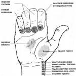
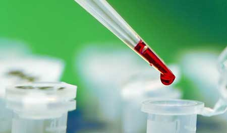
Determination of the quantitative and qualitative composition of blood (hemogram) is carried out, as a rule, by capillary blood, which is taken from a finger with the help of sterile disposable scarification needles and individual sterile pipettes. For biochemical analyzes (more on which will be discussed below) mainly venous blood is used.
Hemoglobin
 Hemoglobin is a red "respiratory" blood pigment. Its main function is the transport function, that is, the transfer of oxygen from the respiratory organs to the tissues, and in the reverse order the transfer of carbon dioxide. Hemoglobin consists of protein (globin) and iron porphyrin (heme), from these two words it got its name. This is the main protein substance of the blood.
Many blood diseases, including hereditary, are associated with impaired hemoglobin structure.
Hemoglobin rates:
- for men - 14.5 g%,
- for women - 13.0 g%.
More broadly, the range of norms depending on gender and age is as follows (g / l):
- newborns - 210;
- infants 2-4 weeks of age - 170.6;
- children aged 1-3 months - 132.6;
- children 4-6 months - 129.2;
- children 7-12 months old - 127.5;
- children from 2 years - 116-135;
- females — 115–145;
- men - 132-164.
If the hemoglobin index is more or less than the limit of the norm, this indicates the presence of pathological conditions. Thus, a decrease in the concentration of hemoglobin in the blood is observed with anemias of various etiologies and with blood loss. This condition is also called anemia. In general, a lack of hemoglobin is already a sign of an anemic condition. As for the types of anemia, there is their classification, formulated by A. I. Vorobiev:
- iron deficiency anemia;
- acute post-hemorrhagic anemia;
- hemolytic anemia;
- anemia associated with inhibition of bone marrow proliferation cells;
- megaloblastic anemia, in which the synthesis of DNA and RNA is impaired;
- sideroachrestrial anemia, in which the porphyrin metabolism is impaired.
An increase in hemoglobin concentration occurs during erythremia (a decrease in the number of red blood cells), erythrocytosis (an increase in the number of red blood cells), as well as when blood is thickened due to a large loss of body fluid. In addition, the hemoglobin index is increased with cardiovascular decompensation.
COLOR INDICATOR
Since hemoglobin is a blood dye, the color indicator expresses the relative content of hemoglobin in one erythrocyte, i.e. the degree of saturation of erythrocytes with hemoglobin. Normally, this degree ranges from 0.85 to 1.15.
The magnitude of the color index is important in determining the form of anemia. Based on the value obtained in the study, anemia is divided into three types:
- hypochromic (color figure is less than 0.85);
- normochromic (color indicator is within the normal range, ie, from 0.85 to 1.15);
- hyperchromic (color index more than 1.15 - the upper limit of the norm).
Erythrocytes and ESR
Red blood cells are red blood cells, or nuclear-free blood cells that contain hemoglobin. They are formed in the bone marrow. The total volume of red blood cells is called the hematocrit value. Knowing this value, we can imagine how the volumes of melt and all the formed elements correlate in the blood.
The normal red blood cell count in men is 4–5 million in 1 μl of blood. Women have fewer of them - “only” 3.7–4.7 million. There is another way to measure the number of red blood cells, where values - namely, their volumes - are expressed in different units of measurement. Thus, the norms of the ratio of blood elements in people who are considered practically healthy look like this.
- Plasma volume - (43.3 + 5.97) ml / kg.
- Red blood cells, volume - (31.8 ± 3.5) ml / kg.
The hematocrit value itself is expressed as a percentage. In men, a normal hematocrit (percentage of red blood cells) is considered to be 40-48%. In women, red blood cells have a slightly smaller proportion, or their share is 36-42%. If the number of erythrocytes is above the norm, then it is usually associated with diseases in which patients have an increased concentration of hemoglobin. The rise in the number of red blood cells is characteristic of:
- any condition of dehydration: toxicosis, vomiting, diarrhea;
- polycytemia;
- adrenal insufficiency;
- congenital heart defects that are associated with cyanosus.
A decrease in the number of erythrocytes is characteristic of an organism with reduced bone marrow function or pathological changes such as leukemia, myeloma, metastasis of malignant tumors, etc.
- hemolytic anemia;
- iron deficiency;
- lack of vitamin B12;
- bleeding.
ESR score
 The determination of erythrocyte sedimentation rate (ESR) is one of the most important and therefore the most commonly prescribed tests. This indicator is expressed in millimeters of plasma, exfoliated within an hour.
The change in ESR is not specific for any disease. However, the acceleration of erythrocyte sedimentation always indicates the presence of a pathological process. As a rule, to assess the processes occurring in the body, the stability of a particular reaction is important. When the pathological process develops, there is a slow acceleration of ESR. After recovery as slowly ESR returns to normal (normal). In women, the ESR indicator is normally from 2 to 14-15 mm / h, for men, from 1 to 10 mm / h.
In children, its rate depends on age and varies as follows:
- 1 mm / h - in newborns;
- 2-6 mm / h - in children up to 1 month;
- 4-14 mm / h - in children from 6 months to 1 year;
- 4-12 mm / h - in children up to 10 years.
Acceleration of ESR, as a rule, is a sign of one of the following conditions of the body:
- infectious diseases;
- inflammatory processes;
- malignant tumors;
- kidney disease;
- liver disease;
- most types of anemia (excluding drepanocytic and microspherocytic anemia);
- anemia associated with impaired protein metabolism, or paraproteinanemia: atypical leukemias, myeloma, macroglobulinemia.
Slowing the ESR and the desire to the lower limit of the norm of this indicator is observed in cardiovascular diseases. One of the reasons for this is an increase in the level of carbon dioxide in the patient's blood.
RETICULOCYTES
Reticulocytes - the name of the particles (formed elements) of blood, relatively little known to a wide readership. Meanwhile, they are young forms of red blood cells. Reticulocytes contain granular inclusions, which scientists have identified using special staining methods. Standards of reticulocyte blood levels are very extensible. Their lower limit is equal to 0.2-1.2%, the upper reaches 12%, which is almost a quarter of all red blood cells in the male body and the third in the female one.
Reticulocytosis - an increase in the blood level of young red blood cells - in humans can be observed in the following cases:
- with anemia;
- malaria;
- in a state of polycemia.
If the number of reticulocytes has fallen and even more so if they have disappeared completely, this is a poor prognostic sign for patients with anemia. This suggests that the bone marrow function of regenerating red blood cells is in a depressed state.
Platelets
Platelets are blood cells that contain the nucleus. They are the smallest in size: their size is only 2-3 microns. They have a big role in the process of blood clotting. Blood clotting is a protective reaction of the body, necessary to prevent blood loss. It should also be noted that the process of blood coagulation is rather complicated; It is regulated by the endocrine and nervous systems.
The opposite of blood clotting quality is fluidity. Normally, the blood has a balanced balance of clotting and fluidity. This is called a hemostatic system. On the one hand, the walls of the vessels themselves (endothelium) secrete substances into the blood, due to which the blood cannot stick together and stick to the walls of the vessels. But, on the other hand, as soon as the vessel is damaged, the substances that form blood clots at the site of damage begin to be released.
During the day, the number of platelets in the blood may vary. In women, it decreases during pregnancy and during menstruation. After physical exertion, platelets become larger than they were at rest. The rate of platelet counts is 180 × 10–– 320 × 109 cells / l. If this figure is less than the norm, doctors say about the so-called thrombocytopenia - a decrease in the level of platelets, which indicates the presence of some disease from the following series:
- hemotyl disease of the newborn;
- acute or (rarely) chronic leukemia;
- chemical poisoning;
- infectious diseases (secondary thrombocytopenia);
- verlgof's disease (primary thrombocytopenia).
In addition, some of the medications taken may lower the platelet count. These are aspirin, sulfonamides, anesthetics and antibiotics. An increase in platelet count is called thrombocytosis and usually occurs in the postoperative period and with:
- asphyxia;
- injuries;
- malignant tumors;
- polycytemia;
- primary idiopathic thrombocythemia.
BLOOD COAGULATION INDICATORS
The bleeding time is determined by its duration from a superficial puncture or incision of the skin. Rate: 1-4 minutes (according to Duke). Clotting time covers the moment from the contact of blood with an alien surface to the formation of a clot. Rate: 6-10 minutes (Lee-White).
In the future, we will return to the topic of blood coagulation and talk about the so-called coagulation factors - special substances that contribute to this process.
White blood cells
Leukocytes are usually called a large group of cells, united under the definition of “white blood cells”. These are colorless blood cells. They are of several types: lymphocytes, monocytes, basophils, eosinophils and neutrophils. All of them have a nucleus and are capable of active amoeboid movement.
The role of leukocytes in our body is huge and very important. They absorb bacteria and dead cells, produce antibodies. These are our cell defenders. Without them, no immunity would be possible and, accordingly, any struggle of the organism against diseases was impossible.
Leukocytes can be found not only in the blood, but also in the lymph. This type of leukocyte is called lymphocyte. By structure, all leukocytes are divided into granular and non-granular. Each type of white blood cells is on guard for the safety of the organism in its own way, that is, it performs its specific functions.
Lymphocytes produce a special type of protein - antibodies that neutralize foreign substances entering the body and their poisons. Some antibodies "work" only against certain substances, others are more versatile - they fight against pathogens of not one, but several diseases. Due to the long-term preservation of antibodies in the body, its overall resistance increases.
Monocytes, they are the phagocytes of the blood (from the Greek phagos - devouring) absorb pathogens, foreign particles, as well as their remnants.
Neutrophils - capable of phagocytosis, as well as monocytes. But their function of body cleaners is even wider: neutrophils destroy viruses, bacteria and their metabolic products - toxins; they detoxify the body, i.e., disinfect it.
Eosinophils - participate in inflammatory processes, allergic reactions, cleansing the body from foreign substances and bacteria. Eosinophils contain antihistamines, which are manifested in allergies.
Basophils - contain histamine and heparin, save the body in case of inflammation and allergic reactions.
The average number of leukocytes ranges from 4 to 9 thousand in 1 μl of blood. The quantitative ratio between the individual forms of leukocytes is called a leukocyte formula. Normally, leukocytes are distributed in the following ratios:
- basophils - 0.1%,
- eosinophils - 0.5-5%,
- stab neutrophils 1-6%,
- segmented neutrophils 47-72%,
- lymphocytes 19-38%
- monocytes 2-11%.
If there are changes in the leukocyte formula, this indicates pathological processes in the body. However, 18 it’s necessary to remember that leukocytosis — an increase in the number of leukocytes in the blood — can be not only pathogenic, but also physiological. Leukocytes increase numerically, for example, during pregnancy. And even active digestion promotes the growth of white blood cells. This does not go beyond the norm. Physiological leukocytosis occurs in healthy people, pathological - in painful conditions.
Causes of physiological leukocytosis:
- food intake (at the same time the number of leukocytes does not exceed 10x10-12 × 109kl / l);
- physical labor;
- hot and cold baths;
- pregnancy;
- childbirth;
- premenstrual period.
By the way, precisely because of the possible distortion of the picture of the analysis due to physiological leukocytosis, blood must be given on an empty stomach. Before “going to the hospital” you shouldn’t do heavy physical work. For pregnant women, parturient women and puerperas their own standards are established. The same applies to children.
Pathological leukocytosis occurs when:
- acute and some chronic infections;
- inflammatory diseases;
- intoxication (nitrobenzene, carbon monoxide, food, quinine, arsenic hydrogen);
- severe oxygen starvation;
- allergic reactions;
- purulent-septic processes;
- malignant tumors;
- blood diseases (leukemia, diseases of the hematopoietic system);
- coma;
- myocardial infarction;
- epilepsy;
- pregnancy at 5-6 months.
Pathological leukocytosis also manifests itself:
- during lactation;
- after heavy blood loss;
- with extensive burns;
- during the premenstrual period;
- after severe physical or mental stress;
- after the introduction of camphor, insulin, adrenaline.
Leukocytosis is usually associated with an increase in the number of neutrophils, less often other types of leukocytes. Thus, the most common causes of pathological leukocytosis are infectious diseases (pneumonia, sepsis, meningitis, pyelonephritis, etc.). Among them can be found infectious diseases with a primary lesion of cells of the immune system (infectious mononucleosis and infectious lymphocytes), as well as various inflammatory diseases caused by microorganisms (peritonitis, phlegmon, etc.). Some infectious diseases always occur with leukopenia. These are typhoid fever, malaria, brucellosis, measles, rubella, flu, viral hepatitis in the acute phase. If there is no leukocytosis in the acute phase of an infectious disease, this is an unfavorable sign, meaning that the body has a weak reactivity (resistance).
The level of leukocytes increases in people suffering from inflammatory diseases of non-microbial etiology, such as, for example, rheumatoid arthritis, systemic lupus erythematosus. The same applies to heart attacks of various organs - myocardium, lungs, etc., since they are based on aseptic (bactericidal) inflammation.
Bone marrow metastases can impair blood formation and cause leukopenia. This is also facilitated by the growth of the body tissue as a result of cell neoplasm, leukemic system blood disease (more than 50 × 10–80 × 109 cells / l of leukocytes) and subleukemic (50 × 10–80 × 10 9 cells / l of leukocytes) forms. When the leukopenic form and aleukemic 20 (the content of leukocytes in the blood is below normal), there will be no forms of leukocytosis.
When the spleen is removed (splenectomy), leukocytosis is observed with an index of 15xU9–20 × 109 cells / l with an increase in the number of neutrophils up to 90%.
But besides leukocytosis may be its opposite. This is leukopenia — a decrease in the number of leukocytes in the blood — which is usually a concomitant symptom:
- radiation damage - exposure to ionizing radiation (X-rays, radiation);
- contact with certain chemicals (benzene, arsenic, DDT, etc.);
- collagenosis (systemic lupus erythematosus);
- taking medications (cytotoxic drugs, some types of antibiotics, sulfonamides, etc.);
- viral and severe bacterial infections;
- diseases of the blood system, in particular leukopenic and aleukemic forms of leukemia, as well as other forms in case of an overdose of cytostatics;
- functional diseases of the central nervous system;
- disorders of blood formation, its insufficiency (bone marrow hypoplasia);
- diseases of the spleen, in which there is increased destruction of blood cells in this organ (cirrhosis of the liver, occurring with an increase in the spleen);
- hodgkin's disease;
- certain endocrine diseases (acromegaly, illness and Cushing's syndrome);
- some infectious diseases (typhoid fever, malaria, influenza, measles, brucellosis, viral hepatitis, prolonged septic endocarditis);
- metastasis of tumors to the bone marrow;
- inflammatory diseases (endometritis, gastritis, colitis, cholecystoangiocholitis) - a lot of leukocytes are eliminated from the body, therefore in severe cases of inflammatory and purulent-septic diseases, the leukocytosis initially replaced by leukopenia).
Often leukopenia is found in old people and malnourished people suffering from inflammatory and purulent-septic diseases. A deficiency of leukocytes is also observed in Addison's disease, sometimes in thyrotoxicosis.
VARIOUS LEUKOCITARIAN FORMULA VIOLATIONS
1. Imbalance in the ratio of neutrophils. Violations of the normal ratio of neutrophils are of several types. The nuclear shift of neutrophils to the left is a condition when many young and degenerative forms of neutrophils appear in the blood. This is usually true for:
- intoxication;
- infectious diseases;
- inflammatory processes;
- malignant tumors.
In this case, there are two types of such a shift - regenerative and degenerative. Regenerative shift - this means that the number of stab and young neutrophils increases on the background of leukocytosis. This suggests an increased activity of the bone marrow, which, as is well known, is the organ of blood formation. This state of the organism is characteristic of purulent-septic and inflammatory processes.
With a degenerative shift, only the number of stab neutrophils increases; while there are degenerative changes in the cells. This suggests that the function of the blood (bone marrow) is depressed.
If at the same time the patient has leukocytosis, then he may have
- toxic dysentery;
- acute peritonitis;
- salmonellosis;
- uremic or diabetic coma.
The degenerative shift of neutrophils on the background of leukopenia speaks about the development:
- immunopathic diseases;
- viral infections.
There is another form of nuclear shift to the left, in which immature forms of leukocytes (myelocytes, promyelocytes or even their predecessors, myeloblasts) appear in the blood. All this happens against the background of a sharp leukocytosis. Such a shift in the blood formula indicates the probable presence of:
- tuberculosis;
- malignant tumors (cancer of the stomach, colon, breast);
- infectious disease.
Professionals know the formula for calculating the severity of the disease by the ratio of leukocytes in the body. Leukocytes in their structure are divided into segmented and non-segmented, each type performs its functions. The ratio of second to first is a value called the “shift index”. This index is calculated by the formula:
shift index = (M + S + P) / C,
in which M is the number of myelocytes, U is the number of young neutrophils, P is the number of stab neutrophils, C is the number of segmented neutrophils.
The normal shift index is expressed in values of 0.05-0.08. Its change in one direction or another indicates the severity of the disease:
- with an index of 1.0 or more - severe;
- in the range of 0.3-1.0 - a disease of moderate severity;
- with an index of 0.3 or less, the degree of the disease is mild.
The neutrophilic nuclear shift to the right is the state of the blood, when neutrophils of mature forms prevail in it, containing five or six instead of three segments. In such cases, the shift index becomes less than the lower limit of the norm — less than 0.04.
For the sake of justice, we should immediately say that the neutrophilic nuclear shift to the right occurs in a fifth of the practically healthy population. However, in some cases it may be a sign of anomalies, in particular, the situation requires further verification due to the suspicion of the presence of:
- radiation sickness;
- polycytemia;
- addisonobirmer anemia.
If the nuclear shift of neutrophils to the right is found in the period of an infectious or inflammatory disease, this is a good sign: the human body actively fights and there is a high probability of a speedy and successful recovery.
2. Other disturbances of leukocyte ratios
Eosinophilia - an increase in the number of eosinophils in the blood. As a rule, this is the body's response to the entry of foreign protein and histamine: after all, these cells perform antihistamine, phagocytic and antitoxic functions. Their increase is typical for the following states:
In contrast to eosinophilia, eosinopenia is a decrease in the number of eosinophils in the blood, and aneosinophilia is their complete absence. These blood conditions are characteristic of diseases such as:
- typhoid fever;
- acute infectious diseases (at the peak of exacerbation);
- agonal condition.
Lymphocytes
Lymphocytes is a type of white blood cell, characterized by its ability to be in the lymph. Their main function is to protect the body from external factors that enter the body in the form of particles of substances and bacteria.
Normally, the absolute content of lymphocytes in the blood should be in the range of 1200-3000 cells / μl. That is, ideally, 1 microliter of blood should contain 1200-3000 lymphocytes.
An increase in the number of lymphocytes above the norm is called lymphocytosis, a decrease is called lymphocytopenia or lymphopenia. Both of these conditions can be absolute and relative. In the first case, the result of the analysis is expressed in the number of cells per unit volume. In the case of relative lymphocytosis or lymphopenia, the analysis data is expressed as a percentage.
As a rule, the change in the number of lymphocytes occurs due to an increase or decrease in the content of other cells in the serum - for example, neutrophils.
Causes of absolute lymphocytosis:
chronic lymphocytic leukemia (proliferative (lat. proles progeny + ferre carry = growth of body tissue as a result of neoplasm of cells) disease of the blood system);
- chronic radiation sickness;
- bronchial asthma;
- thyrotoxicosis (increased thyroid hormone production);
- some infectious diseases (whooping cough, tuberculosis);
- condition after splenectomy (after removal of the spleen);
- taking drugs.
Causes of absolute lymphopenia:
- abnormal development of the lymphoid system (with lymphocyte production in the bone marrow is not enough);
- ionizing radiation (sometimes);
- sometimes - proliferative diseases of the blood system (with leukemia, myeloma, lymphosarcoma, sarcoidosis, carcinoma);
- autoimmune diseases (systemic lupus erythematosus);
- cushing's disease and corticosteroids;
- some forms of tuberculosis (caseous pneumonia, miliary tuberculosis);
- acquired human immunodeficiency syndrome.
T lymphocytes
This is a type of lymphocyte. The cheapest and at the same time sufficiently accurate method for determining the number of T-lymphocytes is the rosetting method. It is based on the presence of affinity between the CD2 T-lymphocyte receptor and glycoproteins (specific antigens) of the sheep erythrocyte membrane. When mixing lymphocytes (serum of the studied blood) with sheep erythrocytes, figures are formed, which are called rosettes. The number of such rosette-forming cells (E-ROCK) corresponds to the number of T-lymphocytes, which are characterized by the presence of antigen on the surface of CD2.
Normally, the relative content of T-lymphocytes is 50–90%, the absolute content is 800–2500 cells / μl, or 0.8 × 10–2.5 × 109 cells / l.
The reasons for the increase in the content of T-lymphocytes:
- diseases of the lymphatic system;
- delayed-type hypersensitivity reactions (DTH) - a type of allergic reaction carried out by T-cells; an example of HRT is allergic dermatitis;
- recovery from the disease when the patient is "recovering";
- tuberculosis.
The reasons for the decrease in the content of T-lymphocytes:
- bacterial chronic infections;
- immunodeficiency;
- tumors;
- tuberculosis;
- stress;
- injury;
- burns;
- hemorrhage;
- some forms of allergies;
- heart attack
T-helpers
Lymphocytes have their own varieties - the so-called subpopulations. Important among them are the so-called T-helpers and T-suppressors. Most often, they are determined with the help of special monoclonal antibodies.
The relative content of these lymphocytes in the amount of 30–50% is considered normal, and the absolute is 600–1600 cells / μl, or 0.6 × 10–1.6 × 109 cells / l.
To determine the ratio between T-helpers and T-suppressors can be in theophylline test. The principle of the method is that in the presence of theophylline substance, T-suppressors lose their ability to E-pink formation. These cells are called those ofillin-sensitive (PM). The so-called theophylline-resistant, i.e., theophylline-resistant cells (TP) in most cases contain T-helpers.
The ratio of TR / PM in the normal range is 2.5-3.5.
The reasons for the increase in the content of T-helper cells:
- infection;
- allergy;
- autoimmune diseases (systemic lupus erythematosus, rheumatoid arthritis, vasculitis, hemolytic anemia, autoimmune glomerulonephritis, etc.).
The reasons for the decrease in the content of T-helper cells:
- immunodeficiency states;
- AIDS;
- cytomegalovirus infection.
Determining the state of T-helper and T-suppressors is included in the immunological study of blood.
Bone marrow irritation often manifests as leukocytosis due to granular (neutrophilic) leukocytes. These changes were systematized by J. Arist and V. Schilling as the study on the "nuclear shift" of neutrophilic leukocytes, which in sufficient detail indicated the degree of changes in bone marrow function. J. Arist subdivided neutrophilic leukocytes into classes and arranged them in the form of a scheme, where the youngest cells were in the extreme graph on the left, and the most mature cells were in the right. The rest maldifferentiated neutrophils are located between them. Subsequently, he included in his classification and other forms of white blood cells. As a result, the scheme became unsuitable because of its bulkiness.
V. Schilling significantly simplified Ariel's scheme, showing that to determine the nuclear shift, only the first class of cells was sufficient for which he introduced the following designations: M - myelocytes, U - young, P - stab and C - segmented neutrophils. When a nuclear shift is taken into account, its nature and direction are noted: a shift to stab-core, to young or to myelocytes. The magnitude of the shift is expressed by a special index (the index of the nuclear shift), which is determined by the formula (M + S + P) / C.
V. Schilling distinguished between regenerative and degenerative shifts of the neutrophil nucleus to the left. Neutrophilic leukocytosis can be with simple hyporegenerative (an increase in the percentage of nuclear leukocytes on the background of neutrophilic leukocytosis), regenerative (an increase in nuclear neutrophils and metamyelocytes - adolescent forms) and hyperregenerative (an increase in neutrophil count up to myelocytes), and regenerative (an increase in the number of neutrophils up to myelocytes), and regenerative (an increase in the number of neutrophils up to myelocytes), and regenerative (an increase in the number of neutrophils up to myelocytes), and regenerative (an increase in the number of neutrophils up to myelocytes), and regenerative (an increase in the number of neutrophils up to myelocytes) nuclei, adrenal glands (young forms) These types of shifts are found in all bone marrow irritation conditions affecting neutrophils, especially in septic diseases.
The degenerative shift proceeds without an increase in the number of leukocytes, with an increase in the percentage of band neutrophils with a change in the structure of the nuclei (pycnotic nuclei), as well as with the appearance of hypersegmented forms of neutrophils, without an increase in the young. A degenerative shift is an expression of histological degeneration, bone marrow suppression, immature, low-producing, cell-poor promyelocytic or myeloblastic bone marrow. It is observed mainly in typhoid fever, less often in tuberculosis and other diseases. In some cases, note the mixed, t. C. regenerative and degenerative shifts to the left. Thus, by “neutrophil nucleus shift”, one can get an idea of the functional activity of the bone marrow.